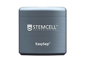
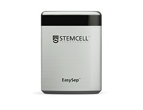
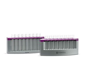
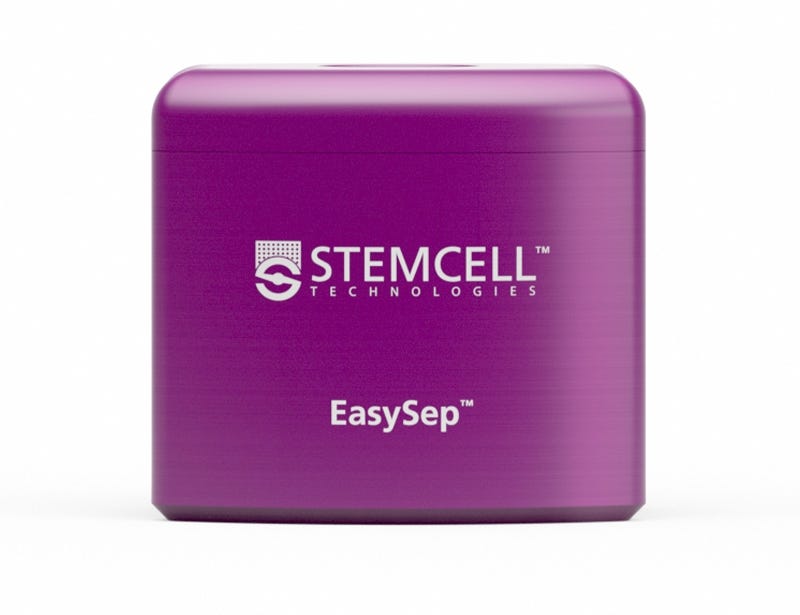
EasySep™ Magnet

Magnet for column-free immunomagnetic separation
New look, same high quality and support! You may notice that your instrument or reagent packaging looks slightly different from images displayed on the website, or from previous orders. We are updating our look but rest assured, the products themselves and how you should use them have not changed. Learn more

EasySep™ Magnet

Magnet for column-free immunomagnetic separation

From: 1,218 USD
Catalog #
(Select a product)
Magnet for column-free immunomagnetic separation
Add to Wish List

What Our Scientist Says

Column-based cell isolation technologies can be complicated, laborious, and time-consuming. We thought there had to be a better way. That's why we developed the column-free EasySep™ cell isolation system.

Jennifer KennettTechnical Scientist
Jennifer Kennett, Technical Scientist

Overview

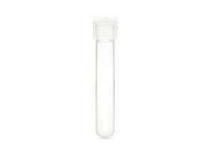
The EasySep™ Magnet is designed for cell separation procedures using EasySep™ reagents. The EasySep™ Magnet generates a high-gradient magnetic field in the interior cavity that is strong enough to separate cells labeled with EasySep™ Magnetic Particles without the use of columns. This magnet is designed to hold a standard 12 x 75 mm (5 mL) polystyrene tube.

Not sure which magnet to use? Visit our EasySep™ Cell Separation Magnets page to compare the different options and select the appropriate magnet for your research.
Species
Human, Mouse, Non-Human Primate, Other, Rat
Application
Cell Isolation
Brand
EasySep

Protocols and Documentation

Find supporting information and directions for use in the Product Information Sheet or explore additional protocols below.

Document Type
Product Name
Catalog #
Lot #
Language
Product Name
EasySep™ Magnet
Catalog #
18000
Lot #
All
Language
English

Applications

This product is designed for use in the following research area(s) as part of the highlighted workflow stage(s). Explore these workflows to learn more about the other products we offer to support each research area.

Research Area
Workflow Stages

Resources and Publications

Educational Materials (18)

Frequently Asked Questions

Can EasySep™ be used for either positive or negative selection?

Yes. The EasySep™ kits use either a negative selection approach by targeting and removing unwanted cells or a positive selection approach targeting desired cells. Depletion kits are also available for the removal of cells with a specific undesired marker (e.g. GlyA).

How does the separation work?

Magnetic particles are crosslinked to cells using Tetrameric Antibody Complexes (TAC). When placed in the EasySep™ Magnet, labeled cells migrate to the wall of the tube. The unlabeled cells are then poured off into a separate fraction.

Which columns do I use?

The EasySep™ procedure is column-free. That's right - no columns!

How can I analyze the purity of my enriched sample?

The Product Information Sheet provided with each EasySep™ kit contains detailed staining information.

Can EasySep™ separations be automated?

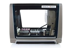
Yes. RoboSep™, the fully automated cell separator, automates all EasySep™ labeling and cell separation steps.

Can EasySep™ be used to isolate rare cells?

Yes. We recommend a cell concentration of 2x10⁸ cells/mL and a minimum working volume of 100 µL. Samples containing 2x10⁷ cells or fewer should be suspended in 100 µL of buffer.

Are the EasySep™ magnetic particles FACS-compatible?

Yes, the EasySep™ particles are flow cytometry-compatible, as they are very uniform in size and about 5000X smaller than other commercially available magnetic beads used with column-free systems.

Can the EasySep™ magnetic particles be removed after enrichment?

No, but due to the small size of these particles, they will not interfere with downstream applications.

Can I alter the separation time in the magnet?

Yes; however, this may impact the kit's performance. The provided EasySep™ protocols have already been optimized to balance purity, recovery and time spent on the isolation.

For positive selection, can I perform more than 3 separations to increase purity?

Yes, the purity of targeted cells will increase with additional rounds of separations; however, cell recovery will decrease.

How does the binding of the EasySep™ magnetic particle affect the cells? is the function of positively selected cells altered by the bound particles?

Hundreds of publications have used cells selected with EasySep™ positive selection kits for functional studies. Our in-house experiments also confirm that selected cells are not functionally altered by the EasySep™ magnetic particles.

If particle binding is a key concern, we offer two options for negative selection. The EasySep™ negative selection kits can isolate untouched cells with comparable purities, while RosetteSep™ can isolate untouched cells directly from whole blood without using particles or magnets.

Publications (5)

Anti-CD105 Antibody Eliminates Tumor Microenvironment Cells and Enhances Anti-GD2 Antibody Immunotherapy of Neuroblastoma with Activated Natural Killer Cells. H.-W. Wu et al. Clinical cancer research : an official journal of the American Association for Cancer Research 2019 may

Abstract

Purpose: We determined whether elimination of CD105+ cells in the tumor microenvironment (TME) with anti-CD105 antibodies enhanced anti-disialoganglioside (GD2) antibody dinutuximab therapy of neuroblastoma when combined with activated natural killer (aNK) cells.Experimental Design: The effect of MSCs and monocytes on antibody-dependent cellular cytotoxicity (ADCC) mediated by dinutuximab with aNK cells against neuroblastoma cells was determined in vitro. ADCC with anti-CD105 mAb TRC105 and aNK cells against MSCs, monocytes, and endothelial cells, which express CD105, was evaluated. Anti-neuroblastoma activity in immunodeficient NSG mice of dinutuximab with aNK cells without or with anti-CD105 mAbs was determined using neuroblastoma cell lines and a patient-derived xenograft.Results: ADCC mediated by dinutuximab with aNK cells against neuroblastoma cells in vitro was suppressed by addition of MSCs and monocytes, and dinutuximab with aNK cells was less effective against neuroblastomas formed with coinjected MSCs and monocytes in NSG mice than against those formed by tumor cells alone. Anti-CD105 antibody TRC105 with aNK cells mediated ADCC against MSCs, monocytes, and endothelial cells. Neuroblastomas formed in NSG mice by two neuroblastoma cell lines or a patient-derived xenograft coinjected with MSCs and monocytes were most effectively treated with dinutuximab and aNK cells when anti-human (TRC105) and anti-mouse (M1043) CD105 antibodies were added, which depleted human MSCs and murine endothelial cells and macrophages from the TME.Conclusions: Immunotherapy of neuroblastoma with anti-GD2 antibody dinutuximab and aNK cells is suppressed by CD105+ cells in the TME, but suppression is overcome by adding anti-CD105 antibodies to eliminate CD105+ cells.
Selective targeting of multiple myeloma by B cell maturation antigen (BCMA)-specific central memory CD8+ cytotoxic T lymphocytes: immunotherapeutic application in vaccination and adoptive immunotherapy. J. Bae et al. Leukemia 2019 mar

Abstract

To expand the breadth and extent of current multiple myeloma (MM)-specific immunotherapy, we have identified various antigens on CD138+ tumor cells from newly diagnosed MM patients (n = 616) and confirmed B-cell maturation antigen (BCMA) as a key myeloma-associated antigen. The aim of this study is to target the BCMA, which promotes MM cell growth and survival, by generating BCMA-specific memory CD8+ CTL that mediate effective and long-lasting immunity against MM. Here we report the identification of novel engineered peptides specific to BCMA, BCMA72-80 (YLMFLLRKI), and BCMA54-62 (YILWTCLGL), which display improved affinity/stability to HLA-A2 compared to their native peptides and induce highly functional BCMA-specific CTL with increased activation (CD38, CD69) and co-stimulatory (CD40L, OX40, GITR) molecule expression. Importantly, the heteroclitic BCMA72-80 specific CTL demonstrated poly-functional Th1-specific immune activities [IFN-gamma/IL-2/TNF-alpha production, proliferation, cytotoxicity] against MM, which were correlated with expansion of Tetramer+ and memory CD8+ CTL. Additionally, heteroclitic BCMA72-80 specific CTL treated with anti-OX40 (immune agonist) or anti-LAG-3 (checkpoint inhibitor) display increased immune function, mainly by central memory CTL. These results provide the framework for clinical application of heteroclitic BCMA72-80 peptide, alone and in combination with anti-LAG3 and/or anti-OX40 therapy, in vaccination and/or adoptive immunotherapeutic strategies to generate long-lasting anti-tumor immunity in patients with MM or other BCMA expressing tumors.
Cold-inducible RNA-binding Protein Induces Neutrophil Extracellular Traps in the Lungs during Sepsis. Y. Ode et al. Scientific reports 2019 apr

Abstract

Extracellular cold-inducible RNA-binding protein (CIRP) exaggerates inflammation and tissue injury in sepsis. Neutrophil extracellular traps (NETs) are released by activated neutrophils during sepsis. NETs contribute to pathogen clearance, but excessive NET formation (NETosis) causes inflammation and tissue damage. Peptidylarginine deiminase 4 (PAD4) is associated with NETosis by increasing histone citrullination and chromatin decondensation. We hypothesized that CIRP induces NETosis in the lungs during sepsis via upregulating PAD4 expression. Sepsis was induced in C57BL/6 wild-type (WT) and CIRP-/- mice by cecal ligation and puncture (CLP). After 20 h of CLP induction, NETs in the lungs of WT and CIRP-/- mice were quantified by flow cytometry by staining the single cell suspensions with MPO and CitH3 Abs. PAD4 expression in the lungs of WT and CIRP-/- mice after sepsis was assessed by Western blotting. In vitro effects of recombinant mouse (rm) CIRP for NETosis and PAD4 expression in the bone marrow-derived neutrophils (BMDN) were assessed by flow cytometry and Western blotting, respectively. After 20 h of CLP, NETosis in the lungs was significantly decreased in CIRP-/- mice compared to WT mice, which also correlated with the decreased PAD4 expression. Intratracheal administration of rmCIRP into WT mice significantly increased NETosis and PAD4 expression in the lungs compared to vehicle-injected mice. In vitro culture of BMDN with rmCIRP significantly increased NETosis and PAD4 expression compared to PBS-treated control. Fluorescence microscopy revealed typical web-like structures consistent with NETs in rmCIRP-treated BMDN. Thus, CIRP serves as a novel inducer of NETosis via PAD4 during sepsis.
New look, same high quality and support! You may notice that your instrument or reagent packaging looks slightly different from images displayed on the website, or from previous orders. We are updating our look but rest assured, the products themselves and how you should use them have not changed. Learn more