Thank you for your interest in this product.
Please provide us with your contact information and your local representative
will contact you with a customized quote. Where appropriate, they can also assist you with a(n):
Estimated delivery time for your area
Product sample or exclusive offer
In-lab demonstration
By submitting this form, you are providing your consent to STEMCELL Technologies Canada Inc. and its subsidiaries and affiliates (“STEMCELL”) to collect and use your information, and send you newsletters and emails in accordance with our privacy policy. Please contact us with any questions that you may have. You can unsubscribe or change your email preferences at any time.
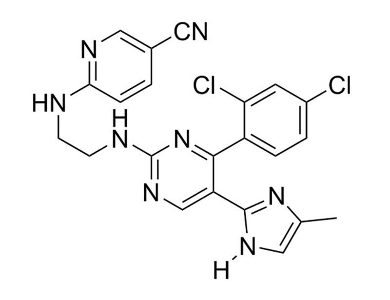
CHIR99021 is an aminopyrimidine derivative that is an extremely potent glycogen synthase kinase (GSK) 3 inhibitor, inhibiting both GSK3β (IC₅₀ = 6.7 nM) and GSK3α (IC₅₀ = 10 nM) (Ring et al.). GSK3 is a serine/threonine kinase that is a key inhibitor of the WNT pathway; therefore CHIR99021 functions as a WNT activator. It shows little activity against a large panel of kinases including CDK2 and other serine/threonine kinases such as MAPK and PKB (Bain et al.).
MAINTENANCE AND SELF-RENEWAL
· Maintains undifferentiated mouse ES cells in combination with PD0325901, in the absence of LIF (Ying et al.).
∙ Promotes self-renewal of human ES cells and mouse epiblast stem cells in combination with IWR-1 (Kim et al.).
∙ Allows derivation of ES cells from refractory mouse strains (Kiyonari et al., Ying et al.) and rat (Li P et al.) in combination with other small molecules.
· Maintains human and mouse hematopoietic stem cells in cytokine-free conditions, in combination with rapamycin (Huang et al.).
· Promotes growth of mouse and human intestinal stem cells (Wang et al.).
REPROGRAMMING
· Enables chemical reprogramming (without genetic factors) of mouse embryonic fibroblasts to iPS cells, in combination with Forskolin, Tranylcypromine, Valproic Acid, 3-Deazaneplanocin A, and E-616452 (Hou et al.).
∙ Promotes reprogramming of human somatic cells to iPS cells using OCT4, in combination with other small molecules (Zhu et al.).
∙ With OCT4, transdifferentiates human CD34+ hematopoietic cells to mesenchymal stem cells (Meng et al.).
· Direct lineage reprogramming of fibroblasts to mature neurons, in combination with Valproic Acid, RepSox, Forskolin, SP600125, Gö6983 and Y-27632 (Hu et al.).
· Direct lineage reprogramming of fibroblasts to mature neurons, in combination with Forskolin, ISX-9, SB431542, and I-BET151 (Li et al.).
· Generates mouse-like or “ground state” iPS cells from human and rat somatic cells, in combination with PD0325901 and A 83-01 (Li W et al. 2009).
DIFFERENTIATION
· Promotes differentiation of insulin-producing cells from human iPS cells (Kunisada et al.).
∙ Promotes differentiation of cardiomyocytes from human ES and iPS cells (Lian et al.).
∙ Generates and maintains primitive neural stem cells from human ES cells, in combination with SB431542 and Human Recombinant LIF (Li W et al. 2011).
This product is designed for use in the following research area(s) as part
of the highlighted workflow stage(s). Explore these workflows to learn more about the other products we
offer to support each research area.
Structural and functional benchmarking of monolayer- and bioreactor-generated hiPSC-derived cardiomyocytes.
Y. Jang et al.
APL bioengineering 2026 Sep
Abstract
Transitioning from animal to human cell sources represents a critical milestone in cardiac tissue engineering and biomedical research. Neonatal rat ventricular myocytes (NRVMs) have long served as the functional benchmark for engineered cardiac tissues; however, their rodent origin limits clinical relevance. Human-induced pluripotent stem cell-derived cardiomyocytes (hiPSC-CMs) offer a renewable, species-specific alternative but remain restricted by immature structure and function, small-scale yield, and high batch variability by conventional two-dimensional monolayer (2D-Mono) differentiation. Here, we systematically evaluated hiPSC-CMs generated by 2D-Mono and three-dimensional embryoid-body (3D-EB) differentiation using identical 15-day Wnt-modulated protocols without additional maturation steps. The 3D-EB method yielded 181 × 106 cells per 100 ml, approximately 2.7-fold higher than the 2D-Mono, while maintaining >80% cTnT+ purity and reduced batch variability. Structural analyses revealed improved sarcomeric organization in 3D-EB tissues compared with 2D-Mono, although both remained less organized than NRVMs, while other morphological parameters were comparable between groups. Functionally, 3D-EB tissues exhibited faster calcium conduction (33.7 cm/s), indicating enhanced electrical coupling relative to 2D-Mono. Although contractile performance remained similar between differentiation formats and below NRVM levels, 3D-EB tissues exhibited consistent structural and functional improvement in calcium wave velocity and contractility over time. These results show that, even without external maturation cues, 3D-EB differentiation yields reproducible, scalable, and human-relevant cardiomyocytes.
Generation of Functional Patient-Specific Thymus Organoids From Human Pluripotent Stem Cells (hPSCs) Using Air–Liquid Interface Culture
S. A. Ramos, H. A. Russ
Bio-protocol 2026 May
Abstract
The thymus is critical for the establishment of a functional and self-tolerant adaptive immune system, but it involutes with age, resulting in reduced naive T-cell output. Generation of a functional human thymus from human pluripotent stem cells (hPSCs) is an attractive regenerative medicine strategy. Direct differentiation of thymic epithelial progenitors (TEPs) from hPSCs has been demonstrated in vitro, but functional thymic epithelial cells (TECs) develop only after transplantation of TEPs in vivo. Functional human reaggregated thymic organoid cultures (RTOCs) and artificial thymic organoids (ATOs) cultured at the air–liquid interface support T-cell development in vitro and in vivo and permit the interrogation of human thymic function and T-cell development. However, these approaches require access to primary human tissues or murine bone marrow stromal cells, are allogeneic, and do not support negative selection. Recently, we reported the directed differentiation of induced PSCs (iPSCs) to functional thymic epithelial progenitors (TEPs) that support murine T-cell development after transplantation in nude mice. Here, we combined hPSC-derived TEPs, hematopoietic progenitor cells (HPCs), and mesenchymal cells, differentiated from the same hPSC line, and generated functional isogenic stem cell–derived thymic organoids (sTOs). Our revised protocol improves our TEP differentiation process and allows the generation of functional isogenic, patient-specific thymic organoids in vitro.
Osimertinib-induced cardiotoxicity is driven by HDAC-dependent epigenetic repression and rescued by vorinostat.
A. Toro Cora et al.
Signal transduction and targeted therapy 2026 Jul
Abstract
Osimertinib, a third-generation EGFR tyrosine kinase inhibitor (TKI), has improved outcomes in non-small cell lung cancer (NSCLC) patients harboring the T790M mutation; however, emerging clinical evidence indicates a risk of cardiotoxicity. Here, we establish the first in vivo preclinical model of osimertinib-induced cardiotoxicity using transverse aortic constriction (TAC) in mice. Osimertinib treatment resulted in cardiac dysfunction, impaired hypertrophic remodeling, and increased markers of heart failure and fibrosis. Unbiased transcriptomic profiling revealed a myocardial stress response characterized by activation of p53-associated cell death pathways, mitochondrial dysfunction, and negative enrichment of histone acetyltransferase (HAT) complexes, indicating epigenetic repression. Mechanistically, osimertinib-treated hearts exhibited increased expression of multiple histone deacetylase (HDAC) isoforms, reduced acetylation of histones, and enhanced cardiomyocyte apoptosis via Bax/caspase-mediated pathways. There was a minimal, transient effect on inflammation, supporting a type I, cell-autonomous cardiotoxic mechanism. Consistent with this, in vitro and in vivo analyses demonstrated suppression of prosurvival ERK/AKT signaling, mitochondrial dysfunction, and activation of intrinsic apoptotic pathways. Given the central role of HDAC activation, we tested whether pharmacologic HDAC inhibition could mitigate osimertinib-induced cardiotoxicity. Treatment with the FDA-approved HDAC inhibitor vorinostat (SAHA) restored histone acetylation, attenuated p53 activation, reduced cardiomyocyte death, and rescued cardiac function in osimertinib-treated mice. Translational studies in human NSCLC-derived PC9 cells further demonstrated that SAHA enhances osimertinib antitumor efficacy while alleviating cardiotoxicity. Collectively, these findings define HDAC-dependent epigenetic repression as a key mechanism underlying osimertinib-induced cardiotoxicity and identify HDAC inhibition as a therapeutically actionable strategy to improve both cardiac safety and cancer treatment efficacy.
PRODUCTS ARE FOR RESEARCH USE ONLY AND NOT INTENDED FOR HUMAN OR ANIMAL DIAGNOSTIC OR THERAPEUTIC USES UNLESS OTHERWISE STATED. FOR ADDITIONAL INFORMATION ON QUALITY AT STEMCELL, REFER TO WWW.STEMCELL.COM/COMPLIANCE.