Human Recombinant EGF

Epidermal growth factor

Human Recombinant EGF

Epidermal growth factor
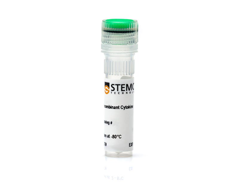
From: 106 USD
Catalog #
(Select a product)
Epidermal growth factor
Add to Wish List

Overview

Epidermal growth factor (EGF) is characterized by high affinity binding to various EGF receptors (EGFRs) and the production of mitogenic responses (Carpenter & Cohen). EGF promotes EGFR dimerization, resulting in activation of downstream pathways including PI3K, ERK1/2, JAK/STAT, β-catenin, and calcium signaling. EGF is secreted by the gut-associated salivary and Brunner’s glands, is found in a variety of body fluids, and stimulates cell proliferation and differentiation in rodent and neonatal human intestine (Wright et al.). Central nervous system stem cells also proliferate in response to the EGF stimulus (Reynolds & Weiss).
Subtype
Cytokines, Growth Factors
Cell Type
Brain Tumor Stem Cells, Endoderm, PSC-Derived, Hematopoietic Stem and Progenitor Cells, Mesenchymal Stem and Progenitor Cells, Mesoderm, PSC-Derived, Neural Cells, PSC-Derived, Neural Stem and Progenitor Cells, Neurons, Pluripotent Stem Cells, Prostate Cells
Species
Human
Area of Interest
Epithelial Cell Biology, Neuroscience, Stem Cell Biology
Purity
> 95%

Data Figures

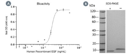
(A) The biological activity of Human Recombinant EGF was tested by its ability to promote the proliferation of BALB/c 3T3 cells. Cell proliferation was measured using a fluorometric assay method. The EC50 is defined as the effective concentration of the growth factor at which cell proliferation is at 50% of maximum. The EC50 in the above example is 0.1 ng/mL.
(B) 2 μg of Human Recombinant EGF was resolved with SDS-PAGE under reducing (+) and non-reducing (-) conditions and visualized by Coomassie Blue staining. Human Recombinant EGF has a predicted molecular mass of 6.2 kDa.

Protocols and Documentation

Find supporting information and directions for use in the Product Information Sheet or explore additional protocols below.

Document Type
Product Name
Catalog #
Lot #
Language
Catalog #
78006.1, 78006, 78006.2
Lot #
All
Language
English
Document Type
Safety Data Sheet
Catalog #
78006.1, 78006, 78006.2
Lot #
All
Language
English

Resources and Publications

Publications (4)

The pro-inflammatory cytokines IFN-α and TNF-α inhibit organoid-derived extravillous trophoblast invasion. A. van Voorden et al. iScience 2026 Jul

Abstract

Proper placental development requires extravillous trophoblast (EVT) differentiation and invasion into the maternal decidua to remodel spiral arteries and support fetal growth. Disruptions in these processes contribute to preeclampsia and fetal growth restriction. These pregnancy complications are common in women with immune-mediated inflammatory diseases, which are characterized by elevated levels of pro-inflammatory cytokines such as interferon-α (IFN-α) and tumor necrosis factor-α (TNF-α). However, the direct effects of these cytokines on EVTs remain unclear. Using human trophoblast organoid models, we demonstrate that IFN-α and TNF-α impair EVT invasion while preserving differentiation capacity. High-resolution imaging of untreated organoid-decidua co-cultures revealed extensive trophoblast invasion into decidual stroma and arteries, but invasion was substantially reduced particularly upon IFN-α treatment. Transcriptome profiling identified changes in several invasion-related pathways after cytokine exposure. These findings suggest that elevated IFN-α and TNF-α can directly impair trophoblast invasive capacity, potentially contributing to suboptimal placental development in inflammatory disorders.
NF-κB and STAT3 signaling uniquely stratify survival in female glioblastoma patients J. Wong et al. iScience 2026 Jan

Abstract

SummaryThe mechanisms underlying sex differences in glioblastoma (GBM) incidence, treatment response, and survival are not well understood. Increased activation of nuclear factor-kappa B (NF-κB) and signal transducer and activator of transcription 3 (STAT3) signaling is associated with shorter survival in GBM. We looked at the expression of NF-κB- or STAT3-related genes in GBM for evidence of a sex skew in activity. Survival analysis of male and female GBM patients revealed that NF-κB- or STAT3-related gene expression was correlated with shorter survival only in female patients. We further explored mechanisms of this sex effect in an established murine model of sex differences in GBM. Concordant with human data, female murine GBM cells expressed stronger signatures of NF-κB and STAT3 genes and exhibited stronger responses to pathway stimulation and inhibition than their male counterparts. This study illustrates the advantage of sex-stratified data analysis in the development of sex-informed treatments for greater precision in cancer treatments. Graphical abstract Highlights•Female GBM patients with upregulated NF-κB/STAT3 signaling have shorter survival•In murine GBM, NF-κB and STAT3 signaling exhibit sex differences•Female murine GBM cells are more sensitive to NF-κB and STAT3 inhibition•Sex-informed use of drugs targeting these pathways could benefit female patients Health disparity; Cancer
FACS-Free isolation and purification protocol of mouse prostate epithelial cells for organoid primary culture. L. Fr\'egeau-Proulx et al. MethodsX 2022

Abstract

The prostate is a gland that contributes to men's fertility. It is highly responsive to androgens and is often the site of carcinogenesis, as prostate cancer is the most frequent cancer in men in over a hundred countries. To study the normal prostate, few in vitro models exist, and most of them do not express the androgen receptor (AR). To overcome this issue, prostate epithelial cells can be grown in primary culture ex vivo in 2- and 3-dimensional culture (organoids). However, methods to purify these cells often require flow cytometry, thus necessitating specialized instruments and expertise. Herein, we present a detailed protocol for the harvest, purification, and primary culture of mouse prostate epithelial cells to grow prostate organoids ex vivo. This protocol does not require flow cytometry approaches, facilitating its implementation in most research laboratories, and organoids grown with this protocol are highly responsive to androgens. In summary, we present a new simple method that can be used to grow prostate organoids that recapitulate the androgen response of this gland in vivo.