STEMdiff™ Intestinal Organoid Kit

Serum-free medium kit for establishment and maintenance of human intestinal organoids from hPSCs
Need a high-quality cell source? Choose from our hiPSC healthy control lines, manufactured with mTeSR™ Plus.

STEMdiff™ Intestinal Organoid Kit

Serum-free medium kit for establishment and maintenance of human intestinal organoids from hPSCs

From: 1,340 USD
Catalog #
(Select a product)
Serum-free medium kit for establishment and maintenance of human intestinal organoids from hPSCs
Add to Wish List

Product Advantages


  • Relevant model system for the developing small intestinal epithelium and associated mesenchyme

  • Robust medium supports efficient differentiation of human ES and iPS cell lines to intestinal organoids

  • Convenient model system that can be maintained long-term through passaging or cryopreserved for experimental flexibility

  • Serum-free medium system optimized for low experimental variability

What's Included

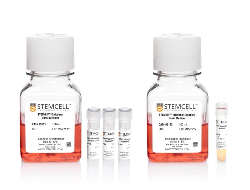
  • STEMdiff™ Endoderm Basal Medium,100 mL
  • STEMdiff™ Definitive Endoderm Supplement CJ, 1.1 mL
  • STEMdiff™ Gastrointestinal Supplement PK, 0.64 mL
  • STEMdiff™ Gastrointestinal Supplement UB, 0.64 mL
  • STEMdiff™ Intestinal Organoid Basal Medium, 100 mL
  • STEMdiff™ Intestinal Organoid Supplement, 2 mL
Products for Your Protocol
To see all required products for your protocol, please consult the Protocols and Documentation.

Overview

STEMdiff™ Intestinal Organoid Kit (Catalog #05140) is a serum-free cell culture medium system that enables robust and efficient generation of human pluripotent stem cell (hPSC)-derived intestinal organoids in a simple, three-stage protocol. Using this kit, hPSCs are directed through induction of definitive endoderm, mid-/hindgut spheroids, and generation of intestinal organoids that can be maintained long-term through passaging or cryopreserved. The organoids exhibit a cellular composition and organization that models the developing intestinal epithelium and associated mesenchyme, making them a convenient model system with direct relevance to the developing intestine. STEMdiff™ Intestinal Organoid Kit is based on the formulation published by Spence et al. (Nature 2011) and has been optimized to increase efficiency and reproducibility of organoid formation and expansion across hPSC lines. Human intestinal organoids can be used as a model system for studying intestinal development and cell biology, intestinal inflammation, intestinal regeneration, microbial interaction, disease modeling, drug discovery, and compound screening. The kit is optimized for differentiation of cells maintained in mTeSR™1, mTeSR™ Plus, or TeSR™-AOF.

For extended culture and passaging, the kit components required for organoid maintenance can be purchased as STEMdiff™ Intestinal Organoid Growth Medium (Catalog #05145).

Browse our Frequently Asked Questions (FAQs) for more information about culturing intestinal organoids using the STEMdiff™ Intestinal Organoid Kit.
Cell Type
Endoderm, PSC-Derived, Intestinal Cells
Species
Human
Application
Cell Culture, Differentiation, Organoid Culture
Brand
STEMdiff
Area of Interest
Disease Modeling, Drug Discovery and Toxicity Testing, Epithelial Cell Biology
Formulation Category
Serum-Free

Data Figures

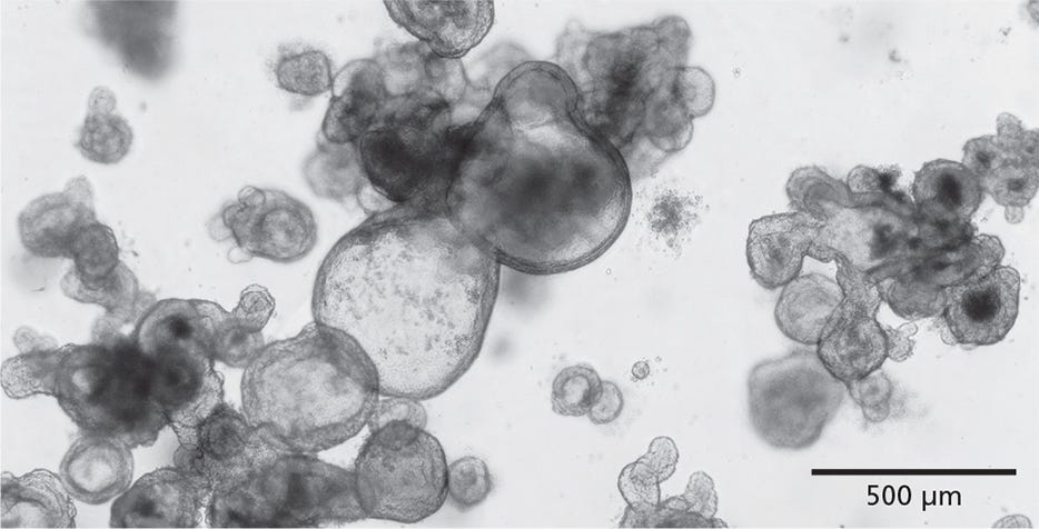
Figure 1. STEMdiff™ Intestinal Organoid Kit Enables the Growth of Intestinal Organoids from PSCs

STEMdiff™ Intestinal Organoid Kit facilitates directed differentiation of PSCs to form human small intestinal organoids. The organoids exhibit a polarized epithelial monolayer surrounding a hollow lumen and are associated with a mesenchymal cell population. Pictured are passage 3 human intestinal organoids generated using this kit.

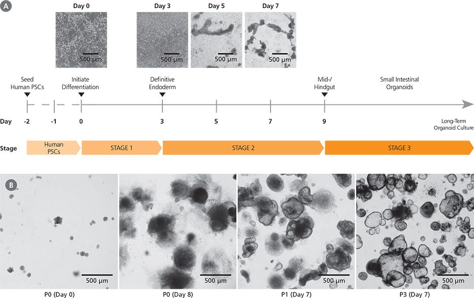
Figure 2. Generation of Human Intestinal Organoid Cultures Using the STEMdiff™ Intestinal Organoid Kit

(A) Human PSC cultures progress through a three-stage differentiation process to generate human intestinal organoids. By day 3 of the protocol, cultures exhibit characteristics typical of definitive endoderm and mid-/hindgut differentiation is initiated. During mid-/hindgut differentiation (days 5 - 9), cells form mid-/hindgut spheroids that are released from the cell monolayer into the culture medium. These spheroids are collected and embedded in extracellular matrix. (B) Embedded mid-/hindgut spheroids cultured in STEMdiff™ Intestinal Organoid Growth Medium mature into intestinal organoids (days in parentheses indicate days post-embedding in a given passage). Once established, intestinal organoids can be maintained and expanded in culture by passaging every 7 - 10 days. After multiple passages, the organoids generally exhibit less sinking within the matrix dome and a lower proportion of mesenchymal cells.

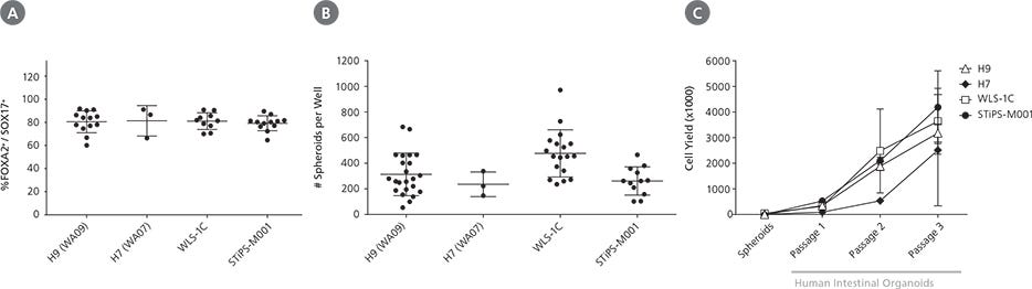
Figure 3. STEMdiff™ Intestinal Organoid Kit Supports Robust Differentiation and Expansion Across ESC and iPSC Lines

STEMdiff™ Intestinal Organoid Kit enables high-efficiency generation of intestinal organoids from both ESCs (H9, H7) and iPSCs (WLS-1C, STiPS-MOO1). (A) Organoids initiated from a variety of cell lines show efficient induction of definitive endoderm, measured by co-expression of FOXA2 and SOX17 on day 3 of differentiation. (B) Both ESCand iPSC-derived cultures demonstrate efficient spheroid formation upon mid-/hindgut induction. The total number of spheroids obtained per well in a given differentiation is shown. (C) Organoids cultured from either ESCs or iPSCs can be expanded and maintained over multiple passages. Shown is the total cell yield per passage. Organoids were passaged every 7 - 10 days with a split ratio between 1 in 2 and 1 in 4. Data points represent the mean of 3 biological replicates. Error bars throughout represent standard deviation of the mean.

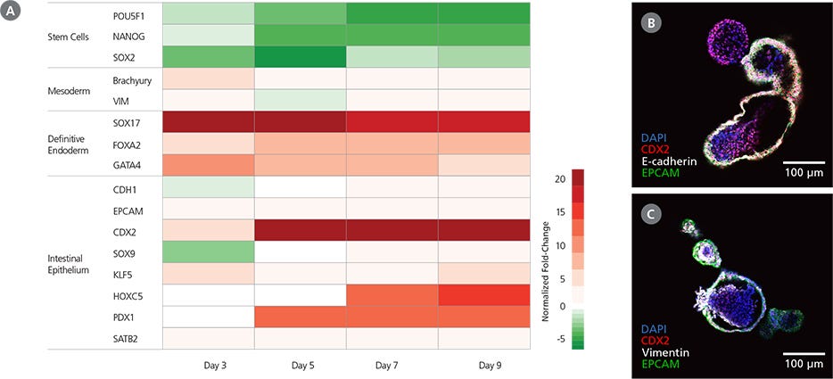
Figure 4. Characteristics of Mid-/Hindgut Spheroids Generated with STEMdiff™ Intestinal Organoid Kit

(A) Cultures differentiated using STEMdiff™ Intestinal Organoid Kit exhibit the expected markers during definitive endoderm and mid-/hindgut specification. During the protocol, gene expression patterns shift from pluripotency markers (day 0) to definitive endoderm markers by day 3 and those of the mid-/hindgut epithelium by day 9. Mid-/hindgut cultures (day 9) also express markers of the associated mesenchyme. Marker levels were assessed by RT-qPCR and normalized to expression levels for undifferentiatied H9 cells. (B) Mid-/hindgut spheroids (day 9) express markers of the intestinal epithelium (CDX2, E-cadherin, EPCAM). (C) Mid-/hindgut spheroids (day 9) also incorporate components of the associated mesenchyme (vimentin).

Figure 5. Intestinal Organoids Cultured In STEMdiff™ Intestinal Organoid Growth Medium Exhibit Features of the Intestinal Epithelium

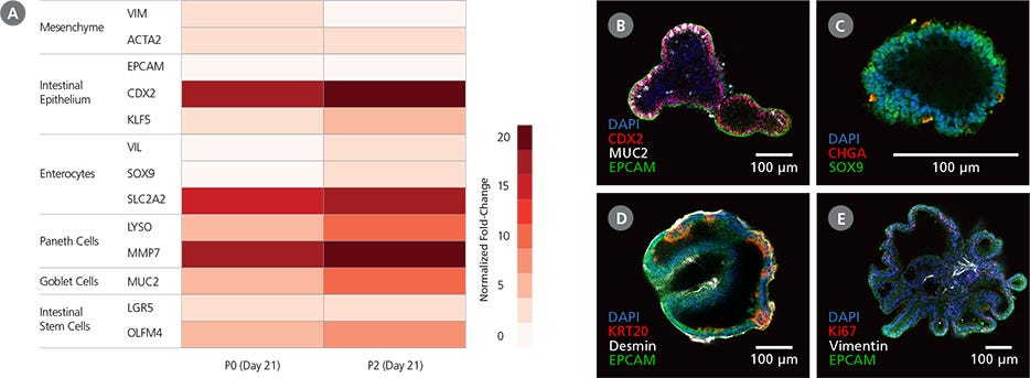
(A) Differentiated PSC-derived intestinal organoids express markers of the intestinal epithelium and the associated mesenchyme. Marker levels were assessed by RT-qPCR and normalized to expression levels for undifferentiated H9 cells. (B,C) Intestinal organoids express markers of intestinal progenitor cells including CDX2 and the intestinal crypt marker SOX9. The organoids are composed of a polarized epithelium, visualized by the localization of EPCAM to the exterior (basolateral) surface of the organoids (B), and express markers typical of mature cell types including MUC2 (B: goblet cells) and CHGA (C: enteroendocrine cells). (D,E) Observation of desmin (D) and vimentin (E) in intestinal organoids demonstrates incorporation of mesenchymal cells in the organoid cultures, while KRT20 (D) and Ki67 (E) are markers of differentiated intestinal cells and putative intestinal stem cells, respectively. Images are digital cross-sections of whole-mount immunofluorescence-stained intestinal organoids at P28 (Day 7).

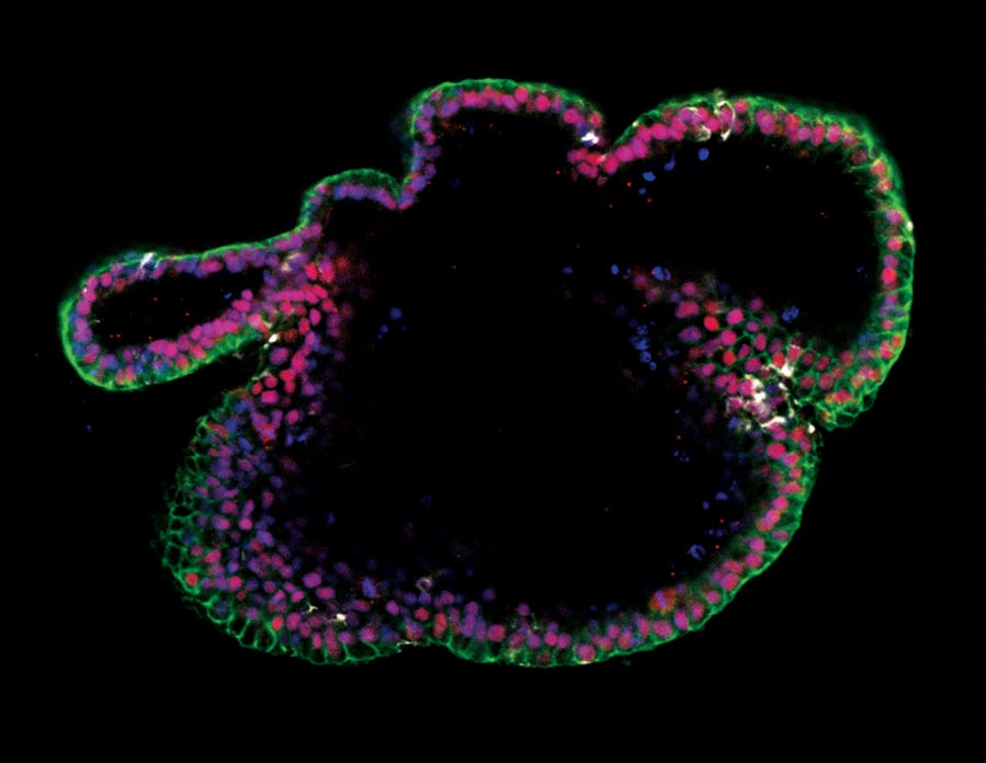
Immunocytochemistry image of an intestinal organoid cultured in mTeSR™ Plus and directed to intestinal organoids using the STEMdiff™ Intestinal Organoid Kit.

Figure 6. Generation of Intestinal Organoids from hPSCs Maintained in mTeSR™ Plus

Human ES (H9) cells were cultured with mTeSR™ Plus and directed to intestinal organoids using the STEMdiff™ Intestinal Organoid Kit. Image shows markers of the intestinal epithelium EpCAM (green) and CDX2 (red), and intestinal mesenchyme marker vimentin (white). Nuclei are counterstained with DAPI (blue).

Protocols and Documentation

Find supporting information and directions for use in the Product Information Sheet or explore additional protocols below.

Document Type
Product Name
Catalog #
Lot #
Language
Catalog #
05140
Lot #
All
Language
English
Document Type
Technical Manual
Catalog #
05140
Lot #
All
Language
English
Document Type
Safety Data Sheet 1
Catalog #
05140
Lot #
All
Language
English
Document Type
Safety Data Sheet 2
Catalog #
05140
Lot #
All
Language
English
Document Type
Safety Data Sheet 3
Catalog #
05140
Lot #
All
Language
English
Document Type
Safety Data Sheet 4
Catalog #
05140
Lot #
All
Language
English
Document Type
Safety Data Sheet 5
Catalog #
05140
Lot #
All
Language
English
Document Type
Safety Data Sheet 6
Catalog #
05140
Lot #
All
Language
English

Applications

This product is designed for use in the following research area(s) as part of the highlighted workflow stage(s). Explore these workflows to learn more about the other products we offer to support each research area.

Resources and Publications

Educational Materials (48)

Publications (7)

Cross-tissue immune profiling of APOE ε4 reveals early dysregulation in Alzheimer's disease. A. Shvetcov et al. Alzheimer's & dementia : the journal of the Alzheimer's Association 2026 Sep

Abstract

INTRODUCTION: Apolipoprotein E (APOE) ε4 is the strongest genetic risk factor for late-onset Alzheimer's disease (AD), but its contribution to disease pathogenesis remains incompletely understood. METHODS: Here, we integrate proteomic profiling of plasma (n = 9028), cerebrospinal fluid (n = 1099), dorsolateral prefrontal cortex (n = 720), and superior temporal gyrus (n = 105) to define the immune phenotype associated with APOE ε4. RESULTS: We identify a conserved, allele dose-dependent pro-inflammatory immune protein signature across peripheral and central tissues independent of AD diagnosis. This signature also emerges in patient-derived cortical organoids prior to amyloid beta and tau pathology, supporting a genotype-driven mechanism. Cross-tissue comparisons reveal shared innate and antiviral responses alongside tissue-specific immune signaling. Notably, a 12-week medical ketogenic diet partially reversed the APOE ε4 immune signature. DISCUSSION: These findings position immune dysregulation as an early and tractable driver of AD risk in APOE ε4 carriers with direct implications for targeted prevention strategies.
IL-22 increases proglucagon-expressing enteroendocrine cells via STAT3 in human iPSC-derived intestinal epithelium. Y. Inoue et al. Endocrinology 2026 Sep

Abstract

Intestinal epithelial differentiation is tightly regulated to maintain tissue homeostasis and systemic metabolic function. Although the intrinsic transcriptional programs governing enteroendocrine cell (EEC) development have been characterized extensively, the role of extrinsic immune signals in directing lineage specification is not completely understood. In particular, how cytokine signaling influences differentiation of EEC subtypes remains unclear. Here, we investigated the role of interleukin-22 (IL-22) in EEC differentiation using human induced pluripotent stem cell-derived intestinal organoids and in vivo models. IL-22 selectively increased proglucagon (GCG) expression and the number of glucagon-like peptide-1 (GLP-1)-positive EECs without affecting other EEC markers. This effect was associated with enhanced expression of the lineage-related transcription factors neurogenin 3 and forkhead box A2. Pharmacological inhibition of signal transducer and activator of transcription 3 (STAT3) signaling attenuated the IL-22-induced increases in GCG expression and GLP-1-positive cell abundance, indicating a role of STAT3 in mediating these effects. Mechanistically, relatively high expression of IL-22 receptor subunit alpha 1 in undifferentiated epithelial cells was associated with STAT3 activation induced by IL-22. Consistent with these findings, in vivo IL-22 administration increased intestinal GCG expression and GLP-1 levels in the circulation and intestinal tissue. Importantly, our findings indicate that IL-22 does not globally promote EEC differentiation but rather selectively directs epithelial lineage commitment toward the GCG-positive EEC fate. These results suggest that IL-22/STAT3 signaling induces human intestinal epithelial differentiation into GCG-positive EECs and may represent a strategy for modulating epithelial cell fate to influence systemic metabolic homeostasis.
Organoid phenotypic screening identified glycyrrhizin that confers protection against tumor necrosis factor-induced cell death Y. Takahashi et al. Stem Cell Reports 2026 Apr

Abstract

Human organoids are considered physiological models that reflect human physiology; however, their applications in drug screening studies are limited. To develop a fundamental treatment for recurrent Crohn’s disease (CD), in which tumor necrosis factor (TNF) is a key pathogenic factor, we conducted phenotypic drug screening to prevent TNF-induced cell death in human intestinal epithelial organoids. Glycyrrhizin, a natural product of licorice root, dose-dependently blocked TNF-induced cell death in organoids but not in TNF-sensitive L929 cells; L929 cells exhibited necroptosis, whereas organoid-derived cells preferentially showed apoptosis upon TNF treatment, determining the specificity of glycyrrhizin. Glycyrrhizin inhibited downstream caspase-8 signaling, which is essential for TNF-dependent apoptosis, and ameliorated intestinal inflammation in vivo. These results demonstrate that glycyrrhizin may be a novel therapeutic compound for CD and highlight the importance of using organoids for phenotypic drug screening. Graphical abstract Highlights•Human intestinal organoids are sensitive to TNF-induced cytotoxicity•We developed an assay system to screen for compounds that resist TNF in organoids•Glycyrrhizin inhibited TNF-induced apoptosis but not necroptosis in organoids•Glycyrrhizin ameliorated intestinal inflammation in a murine model in vivo Takahashi et al. developed a high-throughput phenotypic screening platform using human intestinal organoids, which exhibited higher sensitivity to TNF than conventional intestinal epithelial cell lines, and identified glycyrrhizin that protected TNF-induced cell death both in organoids and in vivo. Phenotypic screening using organoids, as demonstrated in this study, paves the way for drug development and chemical biology.
Need a high-quality cell source? Choose from our hiPSC healthy control lines, manufactured with mTeSR™ Plus.