Anti-Adherence Rinsing Solution

Rinsing solution for cultureware to prevent cell adhesion

Anti-Adherence Rinsing Solution

Rinsing solution for cultureware to prevent cell adhesion
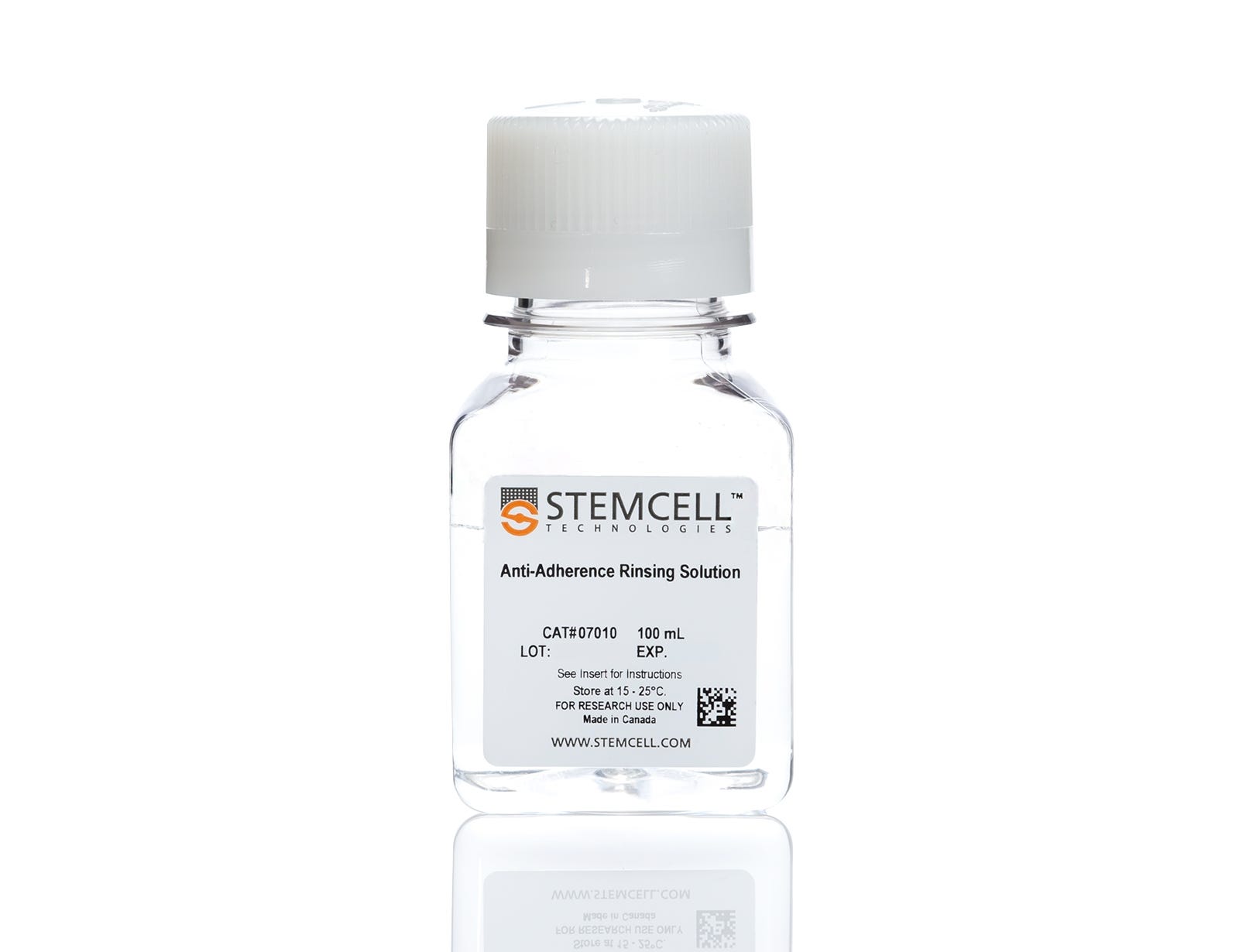
From: 61 USD
Catalog #
(Select a product)
Rinsing solution for cultureware to prevent cell adhesion
Add to Wish List

Overview

Formerly known as AggreWell™ Rinsing Solution, name change effective Dec 2017.

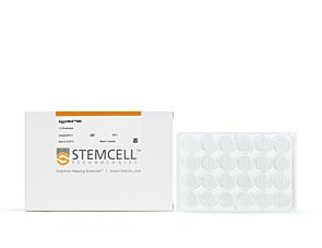
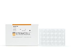
Anti-Adherence Rinsing Solution is a surfactant solution for pre-treating cultureware to reduce surface tension and prevent cell adhesion. It can be used with plates, flasks, pipettes, strainers, and other cultureware.

Anti-Adherence Rinsing Solution is required for use with the following:
• AggreWell™ plates (e.g. Catalog #34811)
• Organoid growth media, including HepatiCult™ (Catalog #06030) and PancreaCult™ (Catalog #06040)
Species
Human, Mouse, Non-Human Primate, Other, Rat
Brand
AggreWell
Area of Interest
Cancer, Neuroscience, Stem Cell Biology

Protocols and Documentation

Find supporting information and directions for use in the Product Information Sheet or explore additional protocols below.

Document Type
Product Name
Catalog #
Lot #
Language
Catalog #
07010
Lot #
All
Language
English
Document Type
Safety Data Sheet
Catalog #
07010
Lot #
All
Language
English

Applications

This product is designed for use in the following research area(s) as part of the highlighted workflow stage(s). Explore these workflows to learn more about the other products we offer to support each research area.

Resources and Publications

Publications (25)

Unconventional auricular reconstruction using controlled scaffold buckling and chondrogenic cocktail containing muscle-derived cells. N. Huang et al. Bioactive materials 2026 Nov

Abstract

Tissue engineered auricles face challenges such as subpar graft mechanical robustness, insufficient stem/progenitor cells, and unidentified factors that specifically promote elastic cartilage. To tackle these, we developed resilient and bioactive 3D-printed scaffolds using two unconventional concepts, including controlled buckling compliant lattices for mechanical resilience and muscle-derived stem/progenitor cells (MDSCs) with defined biochemical factors for facile elastic cartilage-like regeneration. Our functionally graded design reduced stress by 63.1% relative to other graded designs, which was validated by finite element analysis and high-magnitude compressive testing. Notably, 91.7% of scaffolds remained undamaged in stark contrast to 100% failure for clinically-used MEDPOR® and 76.5% failure for other graded scaffolds. Further, we developed an elastic auricular regenerative cocktail (EARc) comprised of abundantly available MDSCs and elastic cartilage-specific biochemical factors. In vitro, mouse, and rabbit studies confirmed that EARc scaffolds facilitated mechanical resilience and elastic cartilage-like regeneration. In conclusion, EARc scaffolds demonstrate the unconventional application of buckling and muscle sourcing to enhance mechanical resilience and elastic-like cartilage-specific regeneration for auricular reconstruction.
Measuring Electrophysiological Activity in Acute Brain Slices, Spheroids, and Organoids Using 3D High-Density Multielectrode Arrays. E. Pali et al. Bio-protocol 2026 Jun

Abstract

Animal and human stem cell-derived three-dimensional models to study physio-pathological brain functioning are becoming a gold standard for in vitro electrophysiology, as they enable the recapitulation of complex network properties by accounting for spatial architectural features that better reflect in vivo conditions than simpler 2D models. Standard planar multielectrode arrays (MEAs), typically providing tens of recording electrodes, are commonly used to record activity from 2D neuronal cultures. However, when adapted for use with 3D models, planar 2D MEAs showed limited effectiveness. The main issues are limited specimen adhesion to the chip, a low number of sensing elements, inability to retrieve signals from within the tissue, and reduced perfusion and vitality of the tissue in contact with sensors. To overcome these limitations, a new generation of microchip-based 3D high-density MEAs (3D HD-MEA) has been developed and validated in recent years. This technological advancement has improved the sensing capabilities and the vitality of 3D models, providing a tool tailored to maximize their potential. Here, we present an optimized protocol for neural network activity recordings in 3D models (including acute slices, brain spheroids, and organoids) from various brain regions using 3D HD-MEAs. First, we summarize the critical steps for 1) obtaining viable acute slices from the mouse cerebellum, cortico-hippocampal circuit, and prefrontal cortex, 2) establishing efficient coupling of the slices with the chip, and 3) performing recordings and analyses. We then describe the main procedures required to obtain human and animal brain spheroids and neural organoids, as well as standardized routines to perform effective recordings and analyses. For each section, we highlight the crucial steps, identify tips for specific applications, and propose troubleshooting procedures. For example, the same type of preparation (e.g., acute slices) requires different adjustments when working with different brain areas. The specific information provided here is intended to assist researchers in their daily efforts to obtain efficient and reproducible functional recordings from 3D models by using the cutting-edge technique of 3D HD-MEA. Key features • Comprehensive all-in-one guide covering the complete workflow for acquiring electrophysiological data from brain slices, neural region-specific organoids, and brain spheroids. • Intuitive, step-by-step protocol for brain slice preparation, enriched with practical tips and expert recommendations to ensure high-quality tissue viability. • Detailed instructions for optimal use of 3D HD-MEA technology, including proper handling of the sample holder for recordings from brain slices, neural organoids, and spheroids. • In-depth guidance on BrainWave6 software, providing clear procedures for data acquisition, signal detection, and advanced electrophysiological analysis across all sample types.
Generation of apical-out nasal organoids to facilitate human respiratory syncytial virus infection and drug screening. G. Stroulios et al. iScience 2026 Jul

Abstract

The nasal epithelium is the first respiratory epithelium that is exposed to inhaled airborne pathogens. As a result, it is crucial to model host-pathogen interactions occurring in this tissue. To facilitate the efficient modeling of these interactions, we have developed a method to generate de novo apical-out nasal organoids from nasal epithelial cell aggregates. Optimization of this method revealed a stark tissue-specific effect of the culture temperature, as organoids were generated in much higher efficiency at 32.5°C compared to more widely used temperatures of 37°C. These organoids recapitulate the native tissue cellular composition of ciliated, basal, and goblet cells, while maintaining high homogeneity in size. Functionally, the system demonstrates susceptibility to viral infection and provides a robust platform for modeling antiviral drug responses. This standardized approach offers a reproducible system with high potential to be utilized in host-pathogen interaction studies and personalized medicine.