How to Prepare Single Cells from PSC-Derived Epithelial Cultures for Flow Cytometry Analysis
The in vitro differentiation of human pluripotent stem cells (hPSCs) into distinct cell and tissue types enables the study of human biology without the need for primary tissues or in vivo models. The STEMdiff™ system provides a standardized procedure for differentiating hPSCs into epithelial cells that can be later cultured in 2D or 3D formats depending on the research needs.
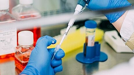
Flow cytometry can be used to measure the efficiency of differentiation protocols with the STEMdiff™ system, by looking at specific marker expression at each stage of the differentiation process. It can also be used for other applications, including cell sorting, immunophenotyping, and purity assessment. Below are protocols for processing and dissociating PSC-derived epithelial cells cultured in different formats, including as 2D monolayers, monolayers in transwell inserts, and organoids, into single cells for downstream analysis and single-cell applications.
Materials
- ACCUTASE™ (Catalog #07920)
- TrypLE™ Express Enzyme (1X), no phenol red (Thermo Fisher Catalog #12604013)
- Gentle Cell Dissociation Reagent (GCDR; Catalog #100-0485)
- FACS buffer
- D-PBS (Without Ca++ and Mg++) (PBS; Catalog #37350)
- 4% paraformaldehyde in PBS (Catalog #100-1450)
- Dulbecco's Phosphate Buffered Saline with 2% Fetal Bovine Serum (Catalog #07905)
- DMEM/F-12 with 15 mM HEPES (Catalog #36254)
*This is a master materials list for the below protocols—all reagents mentioned in each protocol are summarized.
I. Dissociating Monolayers During STEMdiff™ Workflows
This protocol is intended for processing cells generated as monolayers (2D) for flow cytometry assessment as part of the STEMdiff™ differentiation workflow (Table 1).
Table 1. List of STEMdiff™ Kits
The following protocol is for 24-well tissue culture-treated plates. If using an alternate plate format, adjust volumes accordingly (Table 2).
- Rinse wells once with PBS.
- Add 0.5 mL/well of the desired dissociation reagent (e.g. ACCUTASE™ vs TrypLE™) and incubate at 37°C for 5 - 12 minutes. Product-specific guidelines/considerations for incubation are noted below.
- To harvest cells after the definitive endoderm differentiation procedure with STEMdiff™ Definitive Endoderm Kit, we typically recommend incubating with ACCUTASE™ (0.5 mL/well) at 37°C for 5 - 8 minutes.
- If harvesting lung progenitors generated using STEMdiff™ Lung Progenitor Kit, add 0.5 mL of Trypsin-EDTA or TrypLE™ with a final concentration of 0.01 mg/mL DNase I into each well. ACCUTASE™ has not been validated with this workflow.
- If harvesting hepatic progenitors (HPs) or hepatocyte-like cells (HLCs) generated with STEMdiff™ Hepatocyte Kit, incubate with TrypLE™ for 20 minutes at 37°C. ACCUTASE™ has not been validated with this workflow.
Note: Check cells after 5 - 6 minutes of incubation by slightly tapping the plate and checking if monolayer patches lift off; avoid over-digestion.Note: For a very confluent culture, cultures may be a bit more clumpy during dissociation. To overcome this, gently scrape the monolayer with a pipette tip after adding the dissociation reagent to facilitate the dissociation during incubation.Table 2. Volume Adjustment Guidelines for Alternate Plate Formats
96 well-plate50 - 200 μL24 well-plate250 - 500 μL12 well-plate0.5 mL6 well-plate1 mL- Add 1 mL/well of cold FACS buffer to neutralize the dissociation reagent/enzyme.
Notes:- FACS buffer is made up of 2% FBS in sterile PBS (1X PBS).
- EDTA can optionally be added to the FACS buffer at a final concentration of 1 mM.
- If using a different dissociation reagent (e.g. Trypsin-EDTA), add stop medium – 10% FBS in IMDM – to neutralize the reaction.
- It is important to quench the dissociation reagent using FACS buffer or stop medium, by adding at least the same or double the volume of the dissociation reagent.
- Triturate cells with a P1000 pipette to achieve single-cell suspension and transfer to 15 mL conical tubes containing 6 mL of cold FACS buffer.
Notes:- Use pipette tips to gently scratch the bottom of each well in order to lift the monolayer off, then dissociate the cell clumps into a single-cell suspension by pipetting up and down 5 - 10 times.
- Tightly packed monolayers will require some trituration to create single-cell suspensions. Avoid excessive trituration or shear force by resting the pipette tip against the plate edge to maintain viability. If cells do not dissociate well, incubation time with dissociation reagent may need to be optimized.
- For tightly packed pancreatic progenitor cell layers (STEMdiff™ Pancreatic Progenitor Kit), limit trituration to a maximum of 10 cycles (ideally 5). If cells do not dissociate well with trituration, opt for a longer ACCUTASE™ incubation, up to 12 minutes. If monolayers do not dissociate after 12 minutes of ACCUTASE™ incubation and trituration, the differentiation was likely unsuccessful.
- If experiencing low cell viability with ACCUTASE™ when dissociating pancreatic progenitor cell layers (STEMdiff™ Pancreatic Progenitor Kit), incubate monolayers with TrypLE™ at 37°C for 2 - 4 minutes. Check cells after 2 minutes of incubation by slightly tapping the side of the plate or by observing them under the microscope. If cells have not lifted, continue incubation until cells have successfully detached, checking them at 1-minute intervals. Trituration should be performed 3 - 5 times after each incubation. To avoid clumpy cell suspensions, DNAse I can be added at a final concentration of 50 μg/mL to the FACS buffer for the quenching step.
- Going back to the plate, rinse each well with 1 mL of FACS buffer and transfer the volume to the 15 mL tube.
Note: Keep cell suspension on ice after transfer to the tube until ready to run FACS.- Centrifuge for 5 minutes at 300 x g.
- Proceed to antibody staining if performing live cell analysis. Alternatively, proceed to PFA fixation.
Optional protocol for fixing cells with PFA:
Note: PFA fixation could be considered for assessing intracellular markers or performing analysis at a later time.
- Aspirate or decant supernatant from step 6; resuspend in 2 mL/tube of FACS buffer.
- Centrifuge for 5 minutes at 300 x g.
- Resuspend cells in 1 mL/tube of cold 4% PFA in PBS and incubate at room temperature (15 - 25°C) for 10 minutes.Centrifuge for 5 minutes at 300 x g.
- Discard PFA and wash cells in 2 mL/tube of FACS buffer.
- Centrifuge for 5 minutes at 300 x g.
- Resuspend cells in 2 mL/tube of FACS buffer. Fixed cells may be stored at 4°C for up to 2 weeks prior to further analysis.
- The single-cell suspension may now be used for flow cytometry analysis of choice.
II. Dissociating PSC-Derived Epithelial Organoids into Single Cells
The following protocol describes the single-cell dissociation of PSC-derived epithelial organoids. This protocol is only applicable for dome cultures using an extracellular matrix (e.g. Matrigel®). The following protocol is for 24-well tissue culture-treated plates. If using an alternate plate format, adjust volumes accordingly (Table 2).
- Remove the culture medium from the well.
- Dissociate Matrigel® domes using 1 mL of either cold Corning™ Cell Recovery Solution or Cultrex® Organoid Harvesting Solution.
Notes:
- Removal of Matrigel® will be more gentle and efficient for the subsequent dissociation of intestinal organoids into single cells.
- Alternatively, dissociate organoids using Gentle Cell Dissociation Reagent (GCDR) without prior retrieval of organoids from the Matrigel® dome. This will be certainly quicker but bears the risk of insufficient cell dissociation due to issues with Matrigel® and could be more stressful to the cells. When adding GCDR directly to the cultures, pipette up and down multiple times to mechanically break up the dome completely so that GCDR can act on cells for dissociation.
- Cold PBS or DMEM/F-12 could also be used for Matrigel® removal in place of Corning™ Cell Recovery Solution or Cultrex® Organoid Harvesting Solution.
- The incubation is complete when Matrigel® is dissolved and organoids start to float in suspension.
- Once organoids are released from Matrigel® and floating in the harvesting solution, transfer everything into a fresh conical tube. Let organoids settle by gravity or by centrifuging the tube for 5 minutes at 200 x g.
- Carefully remove the supernatant without disturbing the organoid pellet. Add 0.5 mL of desired single-cell dissociation reagent to the tube. Specific incubation guidelines for different dissociation reagents are noted below:
Notes:
- If using ACCUTASE™, an incubation of approximately 8 minutes at 37°C is sufficient to break organoids into single cells.
- If using Gentle Cell Dissociation Reagent (GCDR), the duration of GCDR treatment will have to be optimized. Incubate organoids with GCDR for 8 -10 minutes at 37°C and use a P1000 pipette to gently pipette up and down, and visually inspect if organoids break up. If larger clumps are still visible in the solution, return the tube to 37°C for an additional 2 minutes and repeat the procedure until organoids have completely broken into single cells.
- Alternatively, an incubation with TrypLE™ for 15- 20 minutes could be suggested, depending on how many organoids need to be dissociated into single cells.
- Evaluate the status by checking a droplet and stopping the dissociation when around 80% of the cell suspension is single-celled. Avoid over-digestion.
- Once cells are fully dissociated, add basal media such as DMEM/F-12 or FACS buffer (twice the volume of dissociation reagent used for cell dissociation) to the tube and centrifuge for 5 minutes at 200 x g. Remove supernatant and resuspend cells in FACS buffer.
Note: It is important to quench the dissociation reagent by using the same or double the volume of the dissociation reagent.
- Proceed to antibody staining if performing live cell analysis.
III. Dissociating PSC-Derived Organoids from Transwell® Inserts
The following protocol describes the dissociation of organoids from 24 well-plate Transwell® inserts (Catalog #38024 or Catalog #100-0997) with the STEMdiff™ Branching Lung Organoid Kit. This is an end-point protocol designed to break the branching lung organoids (BLOs) into a single-cell suspension for downstream applications.
- Prepare Dissociation Solution by adding 0.01 mg/mL DNAse to TrypLE™.
- Prepare a Stop Solution by adding 0.01 mg/mL DNAse to FACS buffer (2% FBS and 1mM EDTA in PBS).
Note: It is recommended to include EDTA with FACS buffer for this workflow, as cells often get clumpy and stick together.
- Remove media from apical and basal chambers, being careful not to aspirate BLOs.
- Wash both apical and basal chambers with PBS (300 μL apical, 500 μL basal).
- Add 300 μL and 700 μL Dissociation Solution (prepared in step 1) into apical and basal chambers, respectively.
- Using a P1000 pipette, gently triturate BLOs in the apical chamber approximately 3 times while circling the tip around the insert. Transfer contents to a 15 mL conical tube.
- Repeat steps 5-6 by transferring the Dissociation Solution from the basal to the apical chamber (approximately 4 times).
- Incubate at 37°C for 15 minutes in a water bath.
- Triturate the cell suspension approximately 10 times.
- Inspect 50 μL of cell suspension in a flat-bottom 96-well plate to confirm whether a single-cell suspension has been achieved. Incubate for longer if necessary.
- Add 2 mL Stop Solution to 15 mL falcon tube. Filter the cell suspension (in TrypLE™ + Stop Solution) through cell strainer tubes. This will help remove any residual clumps.
- Centrifuge at 300 x g for 5 minutes.
- Proceed to antibody staining if performing live cell analysis.
IV. Dissociating Kidney Organoids and Underlying Monolayer
The following protocol describes the processing and dissociation of PSC-derived kidney organoids generated using the STEMdiff™ Kidney Organoid Kit. This is an end-point protocol designed to dissociate PSC-derived kidney organoids into a single-cell suspension and fixation for downstream flow cytometry analysis.
- STEMdiff™ Kidney Organoid Kit involves a different plate format/culture set-up and is not a typical monolayer culture; kidney organoids protrude from the underlying monolayer culture.
- This protocol harvests the entire well containing both the kidney organoids and the monolayer.
- This protocol is adapted for 96-well plates; volumes can be adjusted for different plate formats (please refer to Table 1).
- On day 18 of differentiation, wash each well with 200 µL PBS.
- Add 100 μL/well of Collagenase Type I (1 mg/mL) and DNase I Solution (1:500) in HBSS containing Ca++ and Mg++.
- Incubate at 37 °C for 40 minutes.
Note: This time can be optimized. Check for a detached and shriveled-up monolayer. This is indicative of a good dissociation.
- Work quickly at this step to pool wells into a 15 mL tube.
Note: For a large-scale dissociation, use a multichannel pipette to pool cells into a sterile reagent reservoir. Add the pooled wells into a 15 mL tube.Note: For high viability, reduce triturations to less than 5 times before pooling into a tube. If tubules do not dissociate easily, increase collagenase incubation time.
- Centrifuge at 300 x g for 5 minutes.
- Remove supernatant and add ACCUTASE™.
Note: Add 1 mL ACCUTASE™ per 96-well plate. Scale up volumes for increased additional plates.
- Incubate at 37 °C for 6 minutes.
Note: This time can be optimized. Check for small clumps and single cells.
- End dissociation by adding 2 mL FACS buffer. Use a 5 mL pipette to break down any remaining small clumps.
Note: Reduce the number of triturations for higher viability.
- Centrifuge at 300 x g for 5 minutes.
- Remove the supernatant and resuspend the pellet in PBS.
Note: The volume of PBS is dependent on the number of cells harvested.
- Perform cell counts using an automated cell counter.
Note: Low viability can be optimized by the length of collagenase or ACCUTASE™ incubation. It can also be optimized by minimizing the number of triturations.Note: Proceed to antibody staining if performing live cell analysis after this step. Alternatively, proceed to fixable dye staining and PFA fixation.
- Use cell counts to separate the sample into two tubes – “unstained” and “with viability dye”.
- Set the “unstained” tube aside. For the “viability dye” sample, centrifuge the tube at 300 x g for 5 minutes.
- Resuspend in fixable Zombie dye (1:1000) in PBS. Incubate in the dark for 10 minutes.
Note: BioLegend has many options for fixable viability dyes. Choose a dye that will not interfere with any future antibody staining panels. The LIVE/DEAD™ Fixable Near-IR Dead Cell Stain Kit (Thermo) could also be considered.
- Centrifuge both “unstained” and “viability” dye tubes at 300 x g for 5 minutes.
- Aspirate supernatant and add 1 mL of 4% PFA. Incubate for 10-15 minutes at room temperature (15 - 25°C).
- Top up with an equal amount of FACS buffer and centrifuge at 300 x g for 5 minutes.
- Decant and resuspend in 1 mL FACS buffer. Fixed cells may be stored at 4°C for up to 2 weeks prior to further analysis.
- Proceed with analysis.
Additional Resources


Request Pricing
Thank you for your interest in this product. Please provide us with your contact information and your local representative will contact you with a customized quote. Where appropriate, they can also assist you with a(n):
Estimated delivery time for your area
Product sample or exclusive offer
In-lab demonstration

