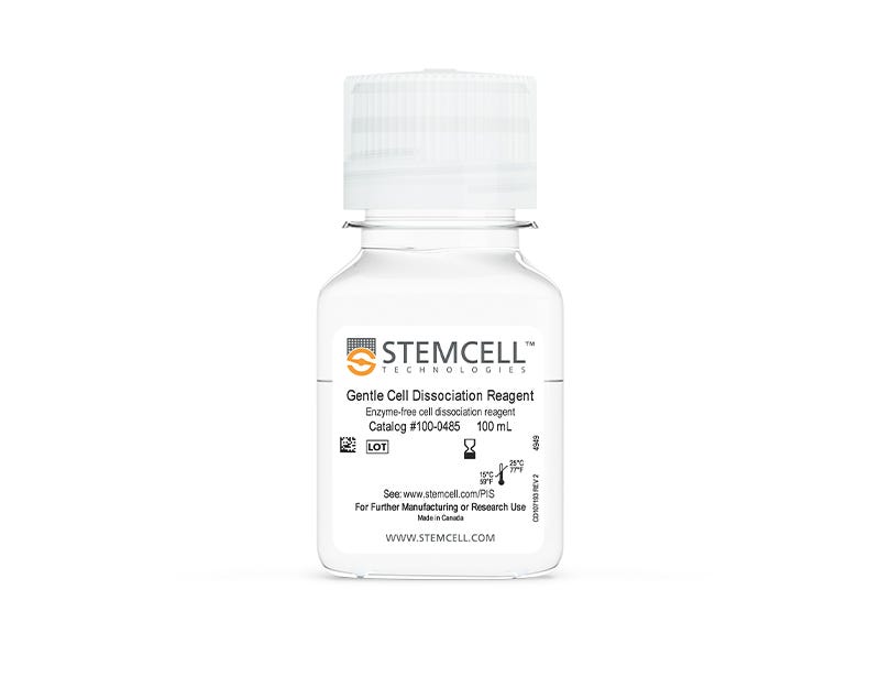
Gentle Cell Dissociation Reagent

cGMP, enzyme-free cell dissociation reagent

Gentle Cell Dissociation Reagent

cGMP, enzyme-free cell dissociation reagent

From: 58 USD
Catalog #
(Select a product)
cGMP, enzyme-free cell dissociation reagent
Add to Wish List

Product Advantages


  • Obtain quality cultures with this gentle, chemically-defined, enzyme-free, GMP solution

  • Achieve high expansion of human ES/iPS cells during routine culture

  • Simplify cell passaging with straightforward, room temperature passaging protocols

Overview

Use Gentle Cell Dissociation Reagent (GCDR) to dissociate human embryonic stem (ES) cells or human induced pluripotent stem (iPS) cells into cell aggregates for routine passaging, or into a single-cell suspension. This enzyme-free and chemically-defined reagent ensures high expansion of human ES and iPS cells during routine culture. It is also suitable for the isolation of intestinal crypts to establish intestinal organoids, and for breaking up Corning® Matrigel® domes when passaging organoid cultures.

GCDR is now also manufactured following relevant cGMPs under a certified quality management system to ensure the highest quality and consistency for reproducible results.

To request a Letter of Authorization (LOA) for Gentle Cell Dissociation Reagent’s Drug Master File, click here.
Subtype
Non-Enzymatic
Cell Type
Endoderm, PSC-Derived, Intestinal Cells, Pluripotent Stem Cells
Species
Human, Mouse
Application
Cell Culture
Area of Interest
Epithelial Cell Biology, Stem Cell Biology

Protocols and Documentation

Find supporting information and directions for use in the Product Information Sheet or explore additional protocols below.

Document Type
Product Name
Catalog #
Lot #
Language
Catalog #
100-0485, 100-1077
Lot #
Lot 1000157161 and lower For 100-0485 | Lot 1000157163 and lower For 100-1077
Language
English
Catalog #
100-0485, 100-1077
Lot #
Lot 1000157162 and higher For 100-0485 | Lot 1000157164 and higher For 100-1077
Language
English
Document Type
Safety Data Sheet
Catalog #
100-0485
Lot #
All
Language
English
Document Type
Safety Data Sheet
Catalog #
100-1077
Lot #
All
Language
English

Resources and Publications

Publications (17)

iPSC-Based Modeling of RAG2 Severe Combined Immunodeficiency Reveals Multiple T Cell Developmental Arrests. M. Themeli et al. Stem cell reports 2020 feb

Abstract

RAG2 severe combined immune deficiency (RAG2-SCID) is a lethal disorder caused by the absence of functional T and B cells due to a differentiation block. Here, we generated induced pluripotent stem cells (iPSCs) from a RAG2-SCID patient to study the nature of the T cell developmental blockade. We observed a strongly reduced capacity to differentiate at every investigated stage of T cell development, from early CD7-CD5- to CD4+CD8+. The impaired differentiation was accompanied by an increase in CD7-CD56+CD33+ natural killer (NK) cell-like cells. T cell receptor D rearrangements were completely absent in RAG2SCID cells, whereas the rare T cell receptor B rearrangements were likely the result of illegitimate rearrangements. Repair of RAG2 restored the capacity to induce T cell receptor rearrangements, normalized T cell development, and corrected the NK cell-like phenotype. In conclusion, we succeeded in generating an iPSC-based RAG2-SCID model, which enabled the identification of previously unrecognized disorder-related T cell developmental roadblocks.
A Novel Toolkit for Characterizing the Mechanical and Electrical Properties of Engineered Neural Tissues. M. Robinson et al. Biosensors 2019 apr

Abstract

We have designed and validated a set of robust and non-toxic protocols for directly evaluating the properties of engineered neural tissue. These protocols characterize the mechanical properties of engineered neural tissues and measure their electrophysical activity. The protocols obtain elastic moduli of very soft fibrin hydrogel scaffolds and voltage readings from motor neuron cultures. Neurons require soft substrates to differentiate and mature, however measuring the elastic moduli of soft substrates remains difficult to accurately measure using standard protocols such as atomic force microscopy or shear rheology. Here we validate a direct method for acquiring elastic modulus of fibrin using a modified Hertz model for thin films. In this method, spherical indenters are positioned on top of the fibrin samples, generating an indentation depth that is then correlated with elastic modulus. Neurons function by transmitting electrical signals to one another and being able to assess the development of electrical signaling serves is an important verification step when engineering neural tissues. We then validated a protocol wherein the electrical activity of motor neural cultures is measured directly by a voltage sensitive dye and a microplate reader without causing damage to the cells. These protocols provide a non-destructive method for characterizing the mechanical and electrical properties of living spinal cord tissues using novel biosensing methods.
Disruption of GRIN2B Impairs Differentiation in Human Neurons. S. Bell et al. Stem cell reports 2018 JUL

Abstract

Heterozygous loss-of-function mutations in GRIN2B, a subunit of the NMDA receptor, cause intellectual disability and language impairment. We developed clonal models of GRIN2B deletion and loss-of-function mutations in a region coding for the glutamate binding domain in human cells and generated neurons from a patient harboring a missense mutation in the same domain. Transcriptome analysis revealed extensive increases in genes associated with cell proliferation and decreases in genes associated with neuron differentiation, a result supported by extensive protein analyses. Using electrophysiology and calcium imaging, we demonstrate that NMDA receptors are present on neural progenitor cells and that human mutations in GRIN2B can impair calcium influx and membrane depolarization even in a presumed undifferentiated cell state, highlighting an important role for non-synaptic NMDA receptors. It may be this function, in part, which underlies the neurological disease observed in patients with GRIN2B mutations.