IntestiCult™ Organoid Growth Medium (Mouse)

Cell culture medium for establishment and maintenance of mouse intestinal organoids
Interested in trying STEMCELL’s organoid products for your intestinal research? Fill out the form to request information about introductory offers.

IntestiCult™ Organoid Growth Medium (Mouse)

Cell culture medium for establishment and maintenance of mouse intestinal organoids

From: 396 USD
Catalog #
(Select a product)
Cell culture medium for establishment and maintenance of mouse intestinal organoids
Add to Wish List

Product Advantages


  • Convenient, in vitro system that recapitulates the identity and organization of the adult intestinal epithelium, including intra- and intercellular signaling, self-propagating stem cell niche and functional transport into and out of the lumen

  • Serum-free and defined medium formulation that delivers consistent results

  • Enables generation of intestinal organoids in less than one week

  • Simple format and easy-to-use protocol

What's Included

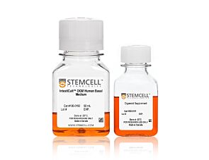
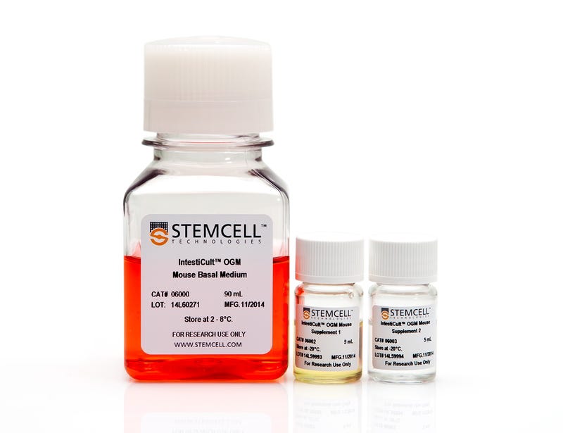
  • IntestiCult™ OGM Mouse Basal Medium, 90 mL
  • IntestiCult™ OGM Mouse Supplement 1, 5 mL
  • IntestiCult™ OGM Mouse Supplement 2, 5 mL
Products for Your Protocol
To see all required products for your protocol, please consult the Protocols and Documentation.

Overview

IntestiCult™ Organoid Growth Medium (Mouse) is a defined, serum-free cell culture medium for efficient establishment and long-term maintenance of mouse intestinal organoids.These organoids, or “mini-guts”, provide a convenient in vitro organotypic culture system for studying both the small and large intestinal epithelium and associated stem cell dynamics. Organoids grown in IntestiCult™ feature a polarized epithelium that contains all of the known cell types of the adult intestinal epithelium. Individual intestinal crypts rapidly form organoids when cultured in IntestiCult™ Organoid Growth Medium (Mouse). Applications of these cultures include studying the development and function of the normal and tumorigenic intestinal epithelium, modeling intestinal disease, and investigating stem cell properties and regenerative therapy approaches. Organoid culture enables convenient in vitro characterization of a system with strong physiological relevance to the adult intestine.

Should you intend to use this product for commercial purposes, please contact HUB at www.huborganoids.nl for a commercial use license or for clarifications in relation to HUB licensing.
Subtype
Specialized Media
Cell Type
Intestinal Cells
Species
Mouse
Application
Cell Culture, Differentiation, Expansion, Maintenance, Organoid Culture
Brand
IntestiCult
Area of Interest
Cancer, Disease Modeling, Drug Discovery and Toxicity Testing, Epithelial Cell Biology, Stem Cell Biology
Formulation Category
Serum-Free

Protocols and Documentation

Find supporting information and directions for use in the Product Information Sheet or explore additional protocols below.

Document Type
Product Name
Catalog #
Lot #
Language
Catalog #
06005
Lot #
All
Language
English
Document Type
Safety Data Sheet 1
Catalog #
06005
Lot #
All
Language
English
Document Type
Safety Data Sheet 2
Catalog #
06005
Lot #
All
Language
English
Document Type
Safety Data Sheet 3
Catalog #
06005
Lot #
All
Language
English

Applications

This product is designed for use in the following research area(s) as part of the highlighted workflow stage(s). Explore these workflows to learn more about the other products we offer to support each research area.

Resources and Publications

Educational Materials (39)

Brochure

Publications (106)

Catechol-O-Methyltransferase Loss Drives Cell-Specific Nociceptive Signaling via the Enteric Catechol-O-Methyltransferase/microRNA-155/Tumor Necrosis Factor ? Axis Q. Zhou et al. Gastroenterology 2023 4

Abstract

BACKGROUND & AIMS The etiology of abdominal pain in postinfectious, diarrhea-predominant irritable bowel syndrome (PI-IBS-D) is unknown, and few treatment options exist. Catechol-O-methyltransferase (COMT), an enzyme that inactivates and degrades biologically active catecholamines, plays an important role in numerous physiologic processes, including modulation of pain perception. Our objective was to determine the mechanism(s) of how decreased colonic COMT in PI-IBS-D patients contributes to the chronic abdominal pain phenotype after enteric infections. METHODS Colon neurons, epithelial cells, and macrophages were procured with laser capture microdissection from PI-IBS-D patients to evaluate cell-specific colonic COMT, microRNA-155 (miR-155), and tumor necrosis factor (TNF) ? expression levels compared to recovered patients (infection cleared: did not develop PI-IBS-D) and control individuals. COMT-/-, colon-specific COMT-/-, and miR-155-/- mice and human colonoids were used to model phenotypic expression of COMT in PI-IBS-D patients and to investigate signaling pathways linking abdominal pain. Citrobacter rodentium and trinitrobenzene sulfonic acid animal models were used to model postinflammatory changes seen in PI-IBS-D patients. RESULTS Colonic COMT levels were significantly decreased and correlated with increased visual analog scale abdominal pain ratings in PI-IBS-D patients compared to recovered patients and control individuals. Colonic miR-155 and TNF-? were increased in PI-IBS-D patients with diminished colonic COMT. COMT-/- mice had significantly increased expression of miR-155 and TNF-? in both colon tissues and dorsal root ganglia. Introduction of cV1q antibody (anti-TNF-?) into mice reversed visceral hypersensitivity after C rodentium and trinitrobenzene sulfonic acid. CONCLUSIONS Decreased colonic COMT in PI-IBS-D patients drives abdominal pain phenotypes via the COMT/miR-155/TNF-? axis. These important findings will allow new treatment paradigms and more targeted and personalized medicine approaches for gastrointestinal disorders after enteric infections.
FUT2-dependent fucosylation of HYOU1 protects intestinal stem cells against inflammatory injury by regulating unfolded protein response. Z. Wang et al. Redox biology 2023 4

Abstract

The intestinal epithelial repair after injury is coordinated by intestinal stem cells (ISCs). Fucosylation catalyzed by fucosyltransferase 2 (FUT2) of the intestinal epithelium is beneficial to mucosal healing but poorly defined is the influence on ISCs. The dextran sulfate sodium (DSS) and lipopolysaccharide (LPS) model were used to assess the role of FUT2 on ISCs after injury. The apoptosis, function, and stemness of ISCs were analyzed using intestinal organoids from WT and Fut2?ISC (ISC-specific Fut2 knockout) mice incubated with LPS and fucose. N-glycoproteomics, UEA-1 chromatography, and site-directed mutagenesis were monitored to dissect the regulatory mechanism, identify the target fucosylated protein and the corresponding modification site. Fucose could alleviate intestinal epithelial damage via upregulating FUT2 and ?-1,2-fucosylation of ISCs. Oxidative stress, mitochondrial dysfunction, and cell apoptosis were impeded by fucose. Meanwhile, fucose sustained the growth and proliferation capacity of intestinal organoids treated with LPS. Contrarily, FUT2 depletion in ISCs aggravated the epithelial damage and disrupted the growth and proliferation capacity of ISCs via escalating LPS-induced endoplasmic reticulum (ER) stress and initiating the IRE1/TRAF2/ASK1/JNK branch of unfolded protein response (UPR). Fucosylation of the chaperone protein HYOU1 at the N-glycosylation site of asparagine (Asn) 862 mediated by FUT2 was identified to facilitate ISCs survival and self-renewal, and improve ISCs resistance to ER stress and inflammatory injury. Our study highlights a fucosylation-dependent protective mechanism of ISCs against inflammation, which may provide a fascinating strategy for treating intestinal injury disorders.
PTBP1 controls intestinal epithelial regeneration through post-transcriptional regulation of gene expression. U. V. Chembazhi et al. Nucleic acids research 2023 3

Abstract

The intestinal epithelial regeneration is driven by intestinal stem cells under homeostatic conditions. Differentiated intestinal epithelial cells, such as Paneth cells, are capable of acquiring multipotency and contributing to regeneration upon the loss of intestinal stem cells. Paneth cells also support intestinal stem cell survival and regeneration. We report here that depletion of an RNA-binding protein named polypyrimidine tract binding protein 1 (PTBP1) in mouse intestinal epithelial cells causes intestinal stem cell death and epithelial regeneration failure. Mechanistically, we show that PTBP1 inhibits neuronal-like splicing programs in intestinal crypt cells, which is critical for maintaining intestinal stem cell stemness. This function is achieved at least in part through promoting the non-productive splicing of its paralog PTBP2. Moreover, PTBP1 inhibits the expression of an AKT inhibitor PHLDA3 in Paneth cells and permits AKT activation, which presumably maintains Paneth cell plasticity and function in supporting intestinal stem cell niche. We show that PTBP1 directly binds to a CU-rich region in the 3' UTR of Phlda3, which we demonstrate to be critical for downregulating the mRNA and protein levels of Phlda3. Our results thus reveal the multifaceted in vivo regulation of intestinal epithelial regeneration by PTBP1 at the post-transcriptional level.
Interested in trying STEMCELL’s organoid products for your intestinal research? Fill out the form to request information about introductory offers.