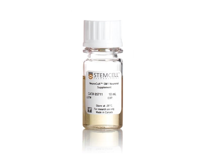
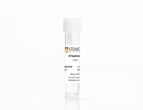
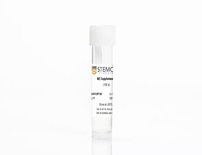
NeuroCult™ SM1 Neuronal Supplement

Supplement (50X) for the serum-free culture of neurons

NeuroCult™ SM1 Neuronal Supplement

Supplement (50X) for the serum-free culture of neurons

From: 121 USD
Catalog #
05711_C
Supplement (50X) for the serum-free culture of neurons

Product Advantages


  • Versatile cell culture supplement

  • Optimized, serum-free formulation

  • Raw materials rigorously screened to maximize lot-to-lot consistency

Overview

Avoid culture failure with a Brewer’s B27-based supplement that provides a consistent neuronal culture experience for CNS-derived or human pluripotent stem cell (hPSC)-derived cells.

NeuroCult™ SM1 (STEMCELL Modified-1) Neuronal Supplement is based on the published formulation (Brewer et al. Brain Res, 1989) and standardized to more reproducibly support survival and maturation of functional neurons. Cultures with this serum-free supplement feature increased neurite outgrowth and branching in short- and long-term cultures. NeuroCult™ SM1 may also be used as a serum-replacement supplement for various customizable applications, such as neurotoxicity assays and calcium imaging.

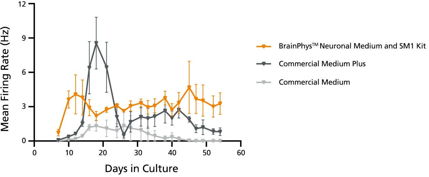
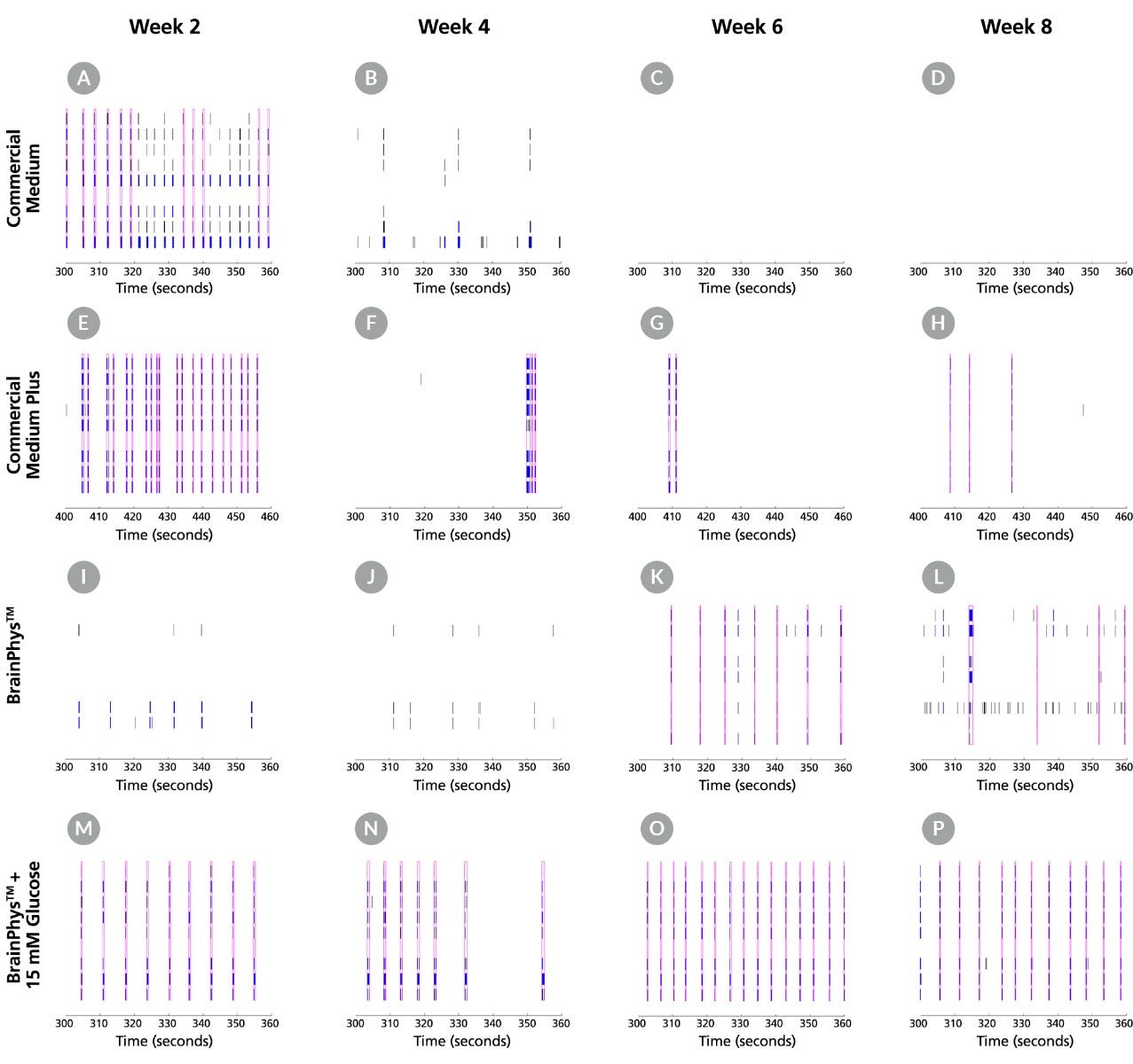
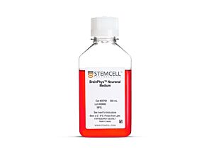
For your convenience, NeuroCult™ SM1 is included as a component of multiple BrainPhys™ Neuronal Medium culture kits for primary and hPSC-derived neurons (Catalog #05792, 05793, 05794, and 05795). For further details, see the performance data with BrainPhys™ below.
Contains
• Antioxidants
• Vitamin A
• Insulin
• Other ingredients
Subtype
Supplements
Cell Type
Neural Cells, PSC-Derived, Neurons, Pluripotent Stem Cells
Species
Human, Mouse, Rat
Application
Cell Culture, Differentiation, Maintenance
Brand
NeuroCult
Area of Interest
Neuroscience, Stem Cell Biology
Formulation
Serum-Free

Data Figures

Morphology of Neurons in Representative NeuroCult™ SM1 Cultures at 7 and 21 Days in Vitro

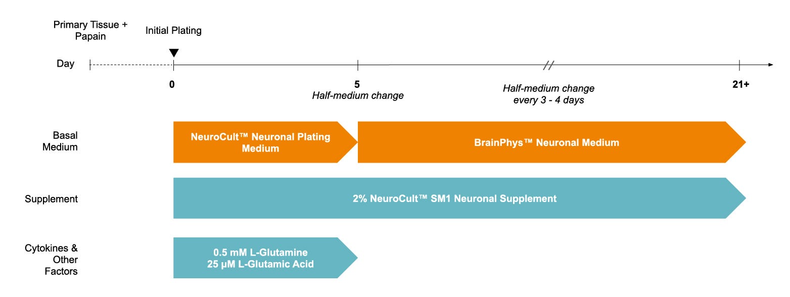
Figure 1. Protocol for Plating and Culturing Primary Neurons with the SM1 Culture System

Primary rodent tissue dissociated in papain was plated in NeuroCult™ Neuronal Plating Medium, supplemented with NeuroCult™ SM1 Neuronal Supplement, L-Glutamine, and L-Glutamic Acid. On day 5, primary neurons were transitioned to BrainPhys™ Neuronal Medium, supplemented with NeuroCult™ SM1 Neuronal Supplement, by performing half-medium changes every 3 - 4 days.

Number of Neurons in NeuroCult™ SM1 and TSFM Cultures After 7 and 21 Days in Vitro

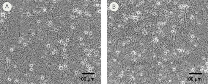
Figure 2. The SM1 Culture System Supports Long-Term Culture of Rodent Neurons

Primary E18 rat cortical neurons were cultured in the SM1 Culture System. A large number of viable neurons are visible after (A) 21 and (B) 35 days, as demonstrated by their bright neuronal cell bodies, and extensive neurite outgrowth and branching. Neurons are evenly distributed over the culture surface with minimal cell clumping.

Neurite Outgrowth of Primary Neurons Cultured in NeuroCult™ SM1 and TSFM for 7 and 21 Days

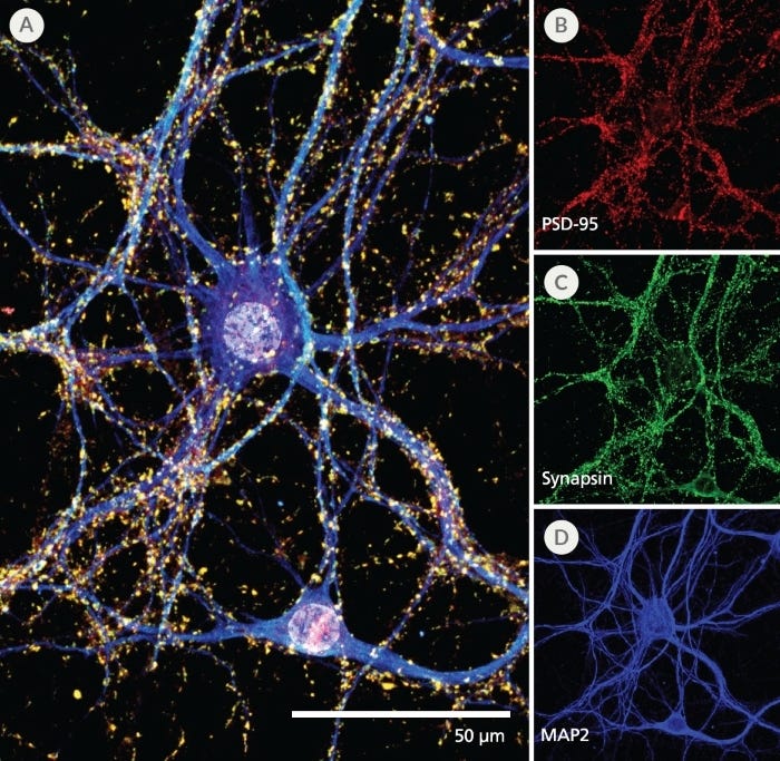
Figure 3. Pre- and Post-Synaptic Markers are Expressed in Rodent Neurons Cultured in the SM1 Culture System

Primary E18 rat cortical neurons were cultured in the SM1 Culture System. At 21 DIV, neurons are phenotypically mature, as indicated by the presence of an extensive dendritic arbor, and appropriate expression and localization of pre-synaptic synapsin (A,C; green) and post-synaptic PSD-95 (A,B; red) markers. Synapsin is concentrated in discrete puncta distributed along the somata and dendritic processes, as defined by the dendritic marker MAP2 (A,D; blue).

Neurite Branching of Primary Neurons Cultured in NeuroCult™ SM1 and TSFM for 7 and 21 Days

Figure 4. The SM1 Culture System Supports Increased Cell Survival

(A) Primary E18 rat cortical neurons were cultured in the SM1 Culture System or a Competitor Culture System for 21 days. Neurons cultured in the SM1 Culture System have a significantly higher number of viable cells compared to the competitor culture system (n = 4; mean ± 95% CI; *p < 0.05). (B) Primary E18 rat cortical neurons were cultured in Neurobasal® supplemented with NeuroCult™ SM1 Neuronal Supplement (SM1) or competitor B27-like supplements (Competitor 1,2,3) for 21 days. Cultures supplemented with NeuroCult™ SM1 Neuronal Supplement have an equal number of neurons compared to competitor-supplemented cultures. Bars represent standard error of mean.

Protocols and Documentation

Find supporting information and directions for use in the Product Information Sheet or explore additional protocols below.

Document Type
Product Name
Catalog #
Lot #
Language
Catalog #
05711
Lot #
All
Language
English
Document Type
Safety Data Sheet
Catalog #
05711
Lot #
All
Language
English

Resources and Publications

Publications (50)

Modelling Lyssavirus Infections in Human Stem Cell-Derived Neural Cultures. V. Sundaramoorthy et al. Viruses 2020 mar

Abstract

Rabies is a zoonotic neurological infection caused by lyssavirus that continues to result in devastating loss of human life. Many aspects of rabies pathogenesis in human neurons are not well understood. Lack of appropriate ex-vivo models for studying rabies infection in human neurons has contributed to this knowledge gap. In this study, we utilize advances in stem cell technology to characterize rabies infection in human stem cell-derived neurons. We show key cellular features of rabies infection in our human neural cultures, including upregulation of inflammatory chemokines, lack of neuronal apoptosis, and axonal transmission of viruses in neuronal networks. In addition, we highlight specific differences in cellular pathogenesis between laboratory-adapted and field strain lyssavirus. This study therefore defines the first stem cell-derived ex-vivo model system to study rabies pathogenesis in human neurons. This new model system demonstrates the potential for enabling an increased understanding of molecular mechanisms in human rabies, which could lead to improved control methods.
Maturation of Human Pluripotent Stem Cell-Derived Cerebellar Neurons in the Absence of Co-culture. T. P. Silva et al. Frontiers in bioengineering and biotechnology 2020

Abstract

The cerebellum plays a critical role in all vertebrates, and many neurological disorders are associated with cerebellum dysfunction. A major limitation in cerebellar research has been the lack of adequate disease models. As an alternative to animal models, cerebellar neurons differentiated from pluripotent stem cells have been used. However, previous studies only produced limited amounts of Purkinje cells. Moreover, in vitro generation of Purkinje cells required co-culture systems, which may introduce unknown components to the system. Here we describe a novel differentiation strategy that uses defined medium to generate Purkinje cells, granule cells, interneurons, and deep cerebellar nuclei projection neurons, that self-formed and differentiated into electrically active cells. Using a defined basal medium optimized for neuronal cell culture, we successfully promoted the differentiation of cerebellar precursors without the need for co-culturing. We anticipate that our findings may help developing better models for the study of cerebellar dysfunctions, while providing an advance toward the development of autologous replacement strategies for treating cerebellar degenerative diseases.
Mutations in ACTL6B Cause Neurodevelopmental Deficits and Epilepsy and Lead to Loss of Dendrites in Human Neurons. S. Bell et al. American journal of human genetics 2019

Abstract

We identified individuals with variations in ACTL6B, a component of the chromatin remodeling machinery including the BAF complex. Ten individuals harbored bi-allelic mutations and presented with global developmental delay, epileptic encephalopathy, and spasticity, and ten individuals with de novo heterozygous mutations displayed intellectual disability, ambulation deficits, severe language impairment, hypotonia, Rett-like stereotypies, and minor facial dysmorphisms (wide mouth, diastema, bulbous nose). Nine of these ten unrelated individuals had the identical de novo c.1027G{\textgreater}A (p.Gly343Arg) mutation. Human-derived neurons were generated that recaptured ACTL6B expression patterns in development from progenitor cell to post-mitotic neuron, validating the use of this model. Engineered knock-out of ACTL6B in wild-type human neurons resulted in profound deficits in dendrite development, a result recapitulated in two individuals with different bi-allelic mutations, and reversed on clonal genetic repair or exogenous expression of ACTL6B. Whole-transcriptome analyses and whole-genomic profiling of the BAF complex in wild-type and bi-allelic mutant ACTL6B neural progenitor cells and neurons revealed increased genomic binding of the BAF complex in ACTL6B mutants, with corresponding transcriptional changes in several genes including TPPP and FSCN1, suggesting that altered regulation of some cytoskeletal genes contribute to altered dendrite development. Assessment of bi-alleic and heterozygous ACTL6B mutations on an ACTL6B knock-out human background demonstrated that bi-allelic mutations mimic engineered deletion deficits while heterozygous mutations do not, suggesting that the former are loss of function and the latter are gain of function. These results reveal a role for ACTL6B in neurodevelopment and implicate another component of chromatin remodeling machinery in brain disease.

Contact STEMCELL Technologies

Our Customer Service, Sales, and Product and Scientific Support departments in North America are available between 6 am and 5 pm Pacific Time (9 am and 8 pm Eastern Time). One of our representatives will be happy to help you by telephone or email. Please complete the form to contact us by email. A representative will get back to you shortly.
  •  

StemCell Technologies Inc. and affiliates ("STEMCELL Technologies") does not share your email address with third parties. StemCell Technologies Inc. will use your email address to confirm your identity and send you newsletters, transaction-related emails, promotional and customer service emails in accordance with our privacy policy. You can change your email preferences at any time.