BrainPhys™ Neuronal Medium

Serum-free neurophysiological basal medium for improved neuronal function
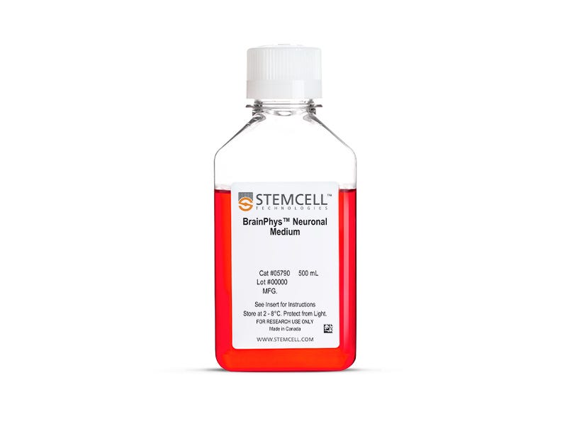
BrainPhys™ Neuronal Medium

Serum-free neurophysiological basal medium for improved neuronal function

From: 136 USD
Catalog #
(Select a product)
Serum-free neurophysiological basal medium for improved neuronal function
Add to Wish List

Product Advantages


  • More representative of the brain’s extracellular environment

  • Improved neuronal function and a higher proportion of synaptically active neurons

  • Perform functional assays without changing media and shocking cells

  • Supports long-term culture of ES/iPS cell- and CNS-derived neurons

  • Rigorous raw material screening and quality control ensure minimal lot-to-lot variability

What Our Scientist Says

I want to help neuroscientists like you create more physiological culture conditions, for more active and healthy neuronal cultures.

Carmen MakScientist
Carmen Mak, Scientist

Overview

Promote, rather than inhibit, neuronal activity and maturity in your cultured primary or human pluripotent stem cell (hPSC)-derived neurons. Based on the formulation by Bardy and Gage (Bardy et al. PNAS, 2015), BrainPhys™ Neuronal Medium is a serum-free basal medium that is optimized to yield a higher proportion of synaptically active neurons by mimicking the central nervous system (CNS) extracellular environment.

Use BrainPhys™ Neuronal Medium for long-term culture of hPSC- and CNS-derived neurons. To avoid shocking your cells with media changes, you can also use BrainPhys™ medium when performing functional assays, such as microelectrode array-based recordings or live-fluorescent imaging.

To ensure cell health in long-term serum-free culture, BrainPhys™ Neuronal Medium must be combined with an appropriate serum-replacement supplement. For researchers culturing hPSC-derived neurons, the BrainPhys™ hPSC Neuron Kit provides BrainPhys™ Neuronal Medium bundled with the recommended supplements for streamlined hPSC differentiation and maturation. For primary neuron cultures, the BrainPhys™ Primary Neuron Kit offers a complete, optimized solution tailored to CNS-derived neurons. If you prefer to customize your workflow, BrainPhys™ Neuronal Medium is also available in convenient kits that include NeuroCult™ SM1 Neuronal Supplement alone (BrainPhys™ Neuronal Medium and SM1 Kit) or in combination with N2 Supplement-A (BrainPhys™ Neuronal Medium N2-A & SM1 Kit).

View our additional resources to learn more about the BrainPhys™ system.
Subtype
Basal Media, Specialized Media
Cell Type
Neural Cells, PSC-Derived, Neurons, Pluripotent Stem Cells
Species
Human, Mouse, Rat
Application
Cell Culture, Differentiation, Maintenance
Brand
BrainPhys
Area of Interest
Disease Modeling, Drug Discovery and Toxicity Testing, Neuroscience, Stem Cell Biology
Formulation Category
Serum-Free

Data Figures

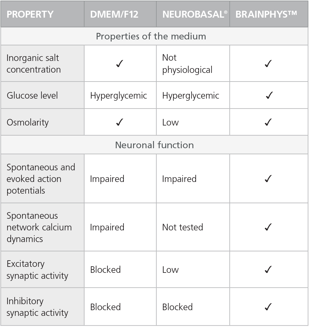
Table 1. Properties of Culture Media (C Bardy et al. Proc Natl Acad Sci USA, 2015)

Check-mark denotes physiological conditions

Check-mark denotes physiological conditions and supported activities according to C Bardy et al. Proc Natl Acad Sci USA, 2015.

Rodent Neurons Matured in BrainPhys™ Neuronal Medium

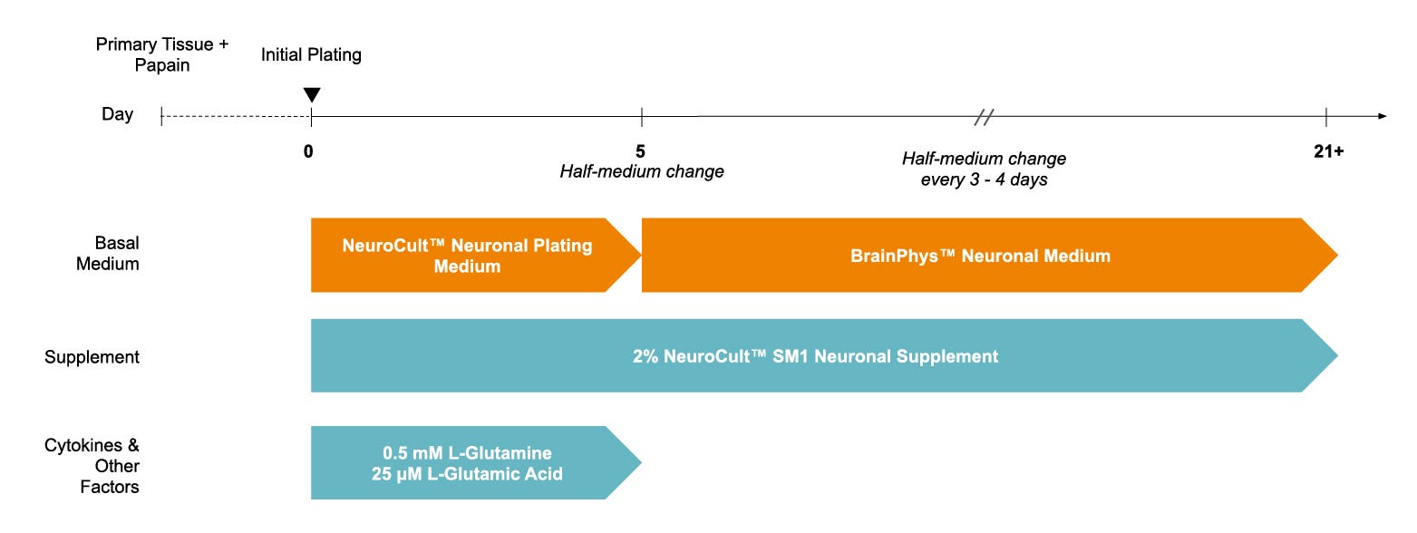
Figure 1. Protocol for Plating and Culturing Primary Neurons with the SM1 Culture System

Primary rodent tissue dissociated in papain was plated in NeuroCult™ Neuronal Plating Medium, supplemented with NeuroCult™ SM1 Neuronal Supplement, L-Glutamine, and L-Glutamic Acid. On day 5, primary neurons were transitioned to BrainPhys™ Neuronal Medium, supplemented with NeuroCult™ SM1 Neuronal Supplement, by performing half-medium changes every 3 - 4 days.

Rodent Neurons Matured in BrainPhys™ Neuronal Medium

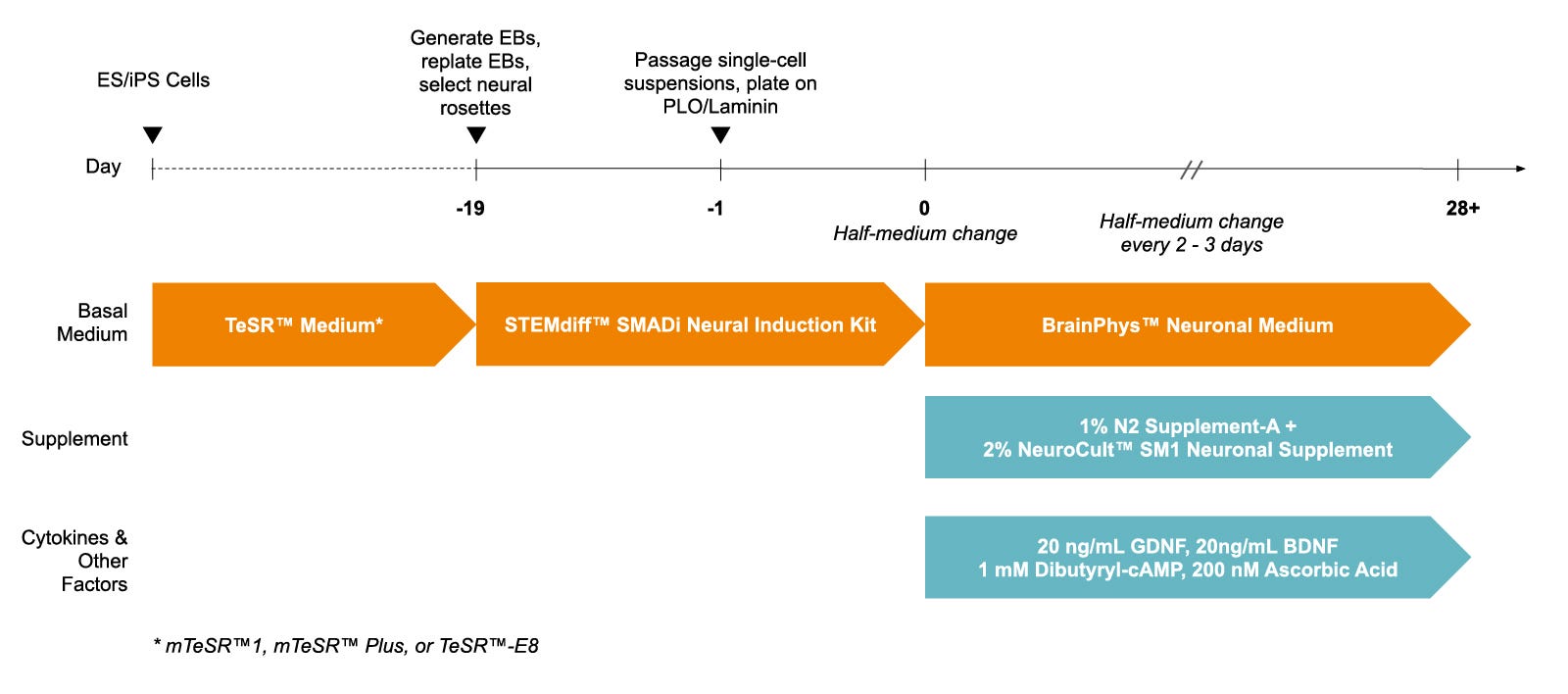
Figure 2. Protocol for Culturing hPSCs with the SM1 Culture System

hPSCs were maintained in mTeSR™1 medium and then differentiated using the STEMdiff™ SMADi Neural Induction Kit. Following plating on PLO/laminin, half-medium changes were performed to transition to BrainPhys™ Neuronal Medium for maturation and long-term culture.

Primary Neuronal Cultures Matured in BrainPhys™ Neuronal Medium Have Greater Numbers of Neurons

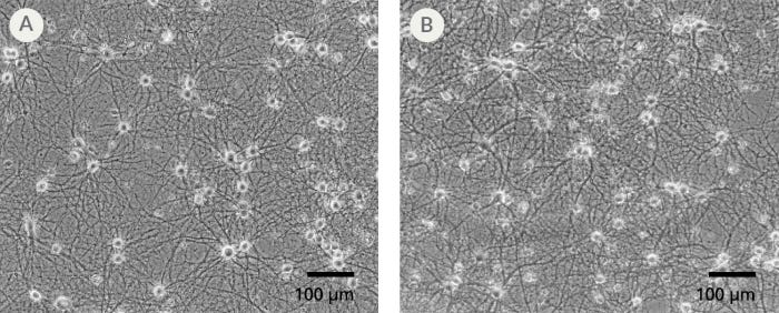
Figure 3. The SM1 Culture System Supports Long-Term Culture of Rodent Neurons

Primary E18 rat cortical neurons were cultured in the SM1 Culture System. A large number of viable neurons are visible after (A) 21 and (B) 35 days, as demonstrated by their bright neuronal cell bodies, and extensive neurite outgrowth and branching. Neurons are evenly distributed over the culture surface with minimal cell clumping.

Rodent Neuronal Cultures Matured in BrainPhys™ Neuronal Medium Show Improved Excitatory and Inhibitory Synaptic Activity

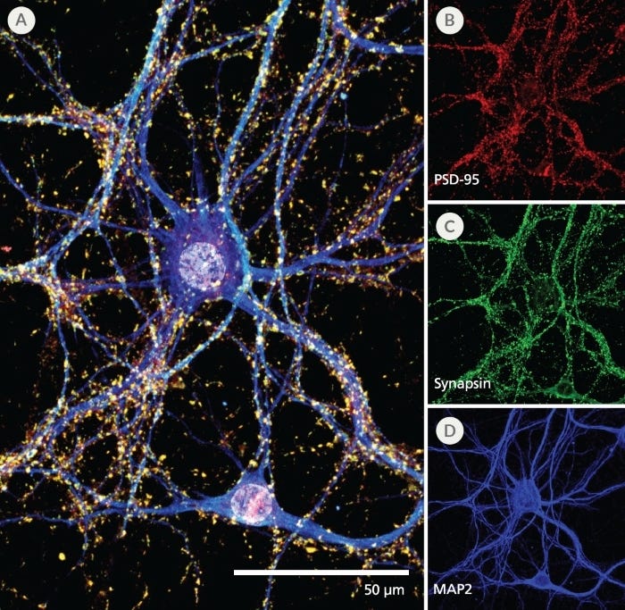
Figure 4. Pre- and Post-Synaptic Markers are Expressed in Rodent Neurons Cultured in the SM1 Culture System

Primary E18 rat cortical neurons were cultured in the SM1 Culture System. At 21 DIV, neurons are phenotypically mature, as indicated by the presence of an extensive dendritic arbor, and appropriate expression and localization of pre-synaptic synapsin (A,C; green) and post-synaptic PSD-95 (A,B; red) markers. Synapsin is concentrated in discrete puncta distributed along the somata and dendritic processes, as defined by the dendritic marker MAP2 (A,D; blue).

Expression of Pre-Synaptic Markers in Rodent Neurons Matured in BrainPhys™ Neuronal Medium

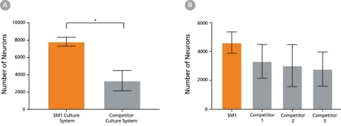
Figure 5. The SM1 Culture System Supports Increased Cell Survival

(A) Primary E18 rat cortical neurons were cultured in the SM1 Culture System or a Competitor Culture System for 21 days. Neurons cultured in the SM1 Culture System have a significantly higher number of viable cells compared to the competitor culture system (n = 4; mean ± 95% CI; *p < 0.05). (B) Primary E18 rat cortical neurons were cultured in Neurobasal® supplemented with NeuroCult™ SM1 Neuronal Supplement (SM1) or competitor B27-like supplements (Competitor 1,2,3) for 21 days. Cultures supplemented with NeuroCult™ SM1 Neuronal Supplement have an equal number of neurons compared to competitor-supplemented cultures. Bars represent standard error of mean.

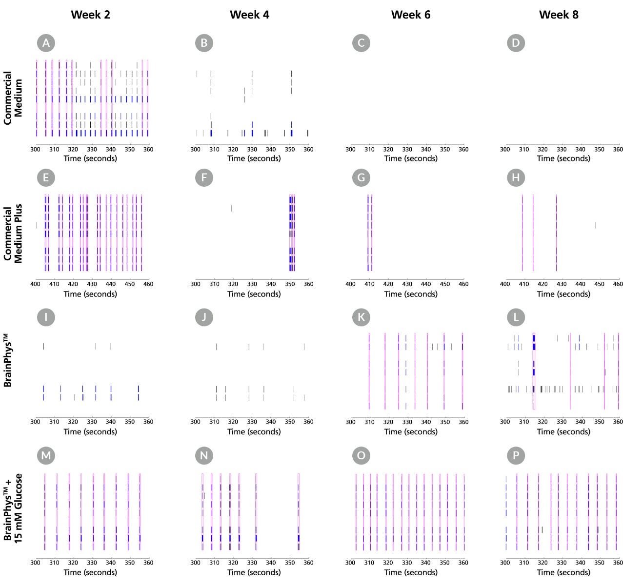
Raster plots showing activity of neurons cultured in BrainPhys and SM1 versus commercial media

Figure 6. BrainPhys™ Supports Improved Neuronal Activity and More Consistent Network Bursting in Long-Term Culture

Raster plots from MEA recordings show the firing patterns of primary E18 rat cortical neurons across 8 electrodes at Weeks 2, 4, 6 and 8. Neurons were either cultured with a Commercial Medium with Supplements, Commercial Medium Plus with Supplements, BrainPhys™ and SM1, or BrainPhys™ and SM1 with 15 mM glucose. Detected spikes (black lines), single channel bursts (blue lines; a collection of at least 5 spikes, each separated by an ISI of no more than 100 ms), and network bursts (magenta boxes; a collection of at least 50 spikes from a minimum of 35% of participating electrodes across each well, each separated by an ISI of no more than 100 ms) were recorded for each medium. (A-D) Neurons cultured with Commercial Medium exhibited network bursting in Week 2 but no spiking activity was detected in subsequent timepoints. (E-H) In Commercial Medium Plus-cultured neurons, a high number of spikes and regular network bursting were detected at Week 2. A decreased number of spikes and inconsistent network bursting were observed in later time points, corresponding to the drop in MFR seen in Figure 4. (I-L) Without glucose, individual spiking was observed at Weeks 2 and 4 with BrainPhys™ and SM1 but network bursting was not detected until Weeks 6 and 8. (M-T) In contrast, neurons cultured with BrainPhys™ and SM1 with 15 mM glucose demonstrated strong spiking activity and consistent network bursting at all timepoints. MEA = microelectrode array; ISI = inter-spike interval; MFR = mean firing rate

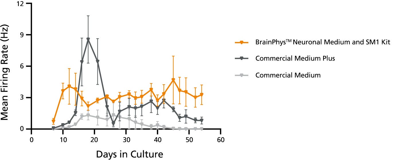
MEA data showing mean firing rate of rodent primary neurons cultured in BrainPhys and SM1 versus commercial media

Figure 7. Glucose Supplementation in BrainPhys™ Maintains Neuronal Activity Over 8 Weeks in Culture

Primary E18 rat cortical neurons were cultured with BrainPhys™ and SM1 or other commercially available culture systems for 8 weeks. Neuronal activity can be detected at Day 9 with BrainPhys™, whereas activity is not detected until Day 14 in cultures maintained in either of the Commercial Media with Commercial Supplements. For Commercial Medium and Supplement-cultured neurons, mean firing rate remains low throughout culture. In contrast, a “peak-drop” activity pattern is observed in the Commercial Medium Plus condition, where mean firing rate increases rapidly within 2 days, followed by a drop in activity in the next 2 - 4 days. BrainPhys™and SM1 Kit with 15 mM glucose maintains the highest level of activity throughout the 8-week culture period.

hPSC-Derived Neurons Generated in BrainPhys™ Neuronal Medium Express Markers of Neuronal Maturity After 14 and 44 Days of Differentiation

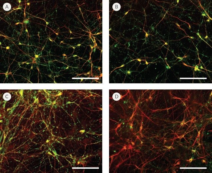
Figure 8. hPSC-Derived Neurons Generated in BrainPhys™ Neuronal Medium Express Markers of Neuronal Maturity After 14 and 44 Days of Differentiation

NPCs were generated from H9 cells using STEMdiff™ Neural Induction Medium in an embryoid body-based protocol. Next, NPCs were cultured in (A,C) BrainPhys™ Neuronal Medium, supplemented with 2% NeuroCult™ SM1 Supplement, 1% N2 Supplement-A, 20 ng/mL GDNF, 20 ng/mL BDNF, 1 mM db-cAMP and 200 nM ascorbic acid to initiate neuronal differentiation, or (B,D) DMEM/F12 under the same supplementation conditions. After 14 and 44 days of differentiation and maturation, neurons express the synaptic marker Synapsin 1 (green) and the mature neuronal marker MAP2 (red). In this example, neurons matured in BrainPhys™ Neuronal Medium show increased Synapsin 1 staining. Scale bar= 100 µm

hPSC-Derived Neurons Generated in BrainPhys™ Neuronal Medium and NeuroCult™ SM1 and N2 Supplements are Healthy and Morphologically Normal

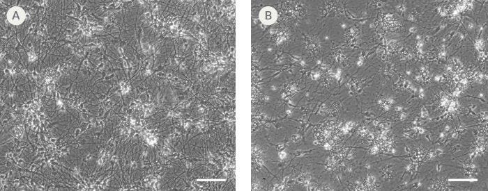
Figure 9. hPSC-Derived Neurons Generated in BrainPhys™ Neuronal Medium and NeuroCult™ SM1 and N2 Supplements are Healthy and Morphologically Normal

NPCs were generated from H9 cells using STEMdiff™ Neural Induction Medium in an embryoid body-based protocol. Next, NPCs were cultured for 44 DIV in (A) BrainPhys™ Neuronal Medium, supplemented with 2% NeuroCult™ SM1 Supplement, 1% N2 Supplement-A, 20 ng/mL GDNF, 20 ng/mL BDNF, 1 mM db-cAMP and 200 nM ascorbic acid to initiate neuronal differentiation, or (B) DMEM/F12 under the same supplementation conditions. Neuronal cultures differentiated from NPCs in BrainPhys™ Neuronal Medium display extensive neurite outgrowth and reduced cellular debris compared to cultures differentiated in DMEM/F12. Scale bar= 100 µm.

hPSC-Derived Neurons Matured in BrainPhys™ Neuronal Medium Show Improved Excitatory and Inhibitory Synaptic Activity

Figure 10. hPSC-Derived Neurons Matured in BrainPhys™ Neuronal Medium Show Improved Excitatory and Inhibitory Synaptic Activity

NPCs were generated from H9 cells using STEMdiff™ Neural Induction Medium in an embryoid body-based protocol. Next, NPCs were cultured for 44 DIV in (A,C) BrainPhys™ Neuronal Medium, supplemented with 2% NeuroCult™ SM1 Supplement, 1% N2 Supplement-A, 20 ng/mL GDNF, 20 ng/mL BDNF, 1 mM db-cAMP and 200 nM ascorbic acid to initiate neuronal differentiation, or (B,D) in DMEM/F12 under the same supplementation conditions. (A,C) Neurons matured in BrainPhys™ Neuronal Medium showed spontaneous excitatory (AMPA-mediated; A) and inhibitory (GABA-mediated; C) synaptic events. The frequency and amplitude of spontaneous synaptic events is consistently greater in neuronal cultures matured in BrainPhys™ Neuronal Medium, compared to neurons plated and matured in DMEM/F12 (B,D). Traces are representative.

Protocols and Documentation

Find supporting information and directions for use in the Product Information Sheet or explore additional protocols below.

Document Type
Product Name
Catalog #
Lot #
Language
Catalog #
05790
Lot #
All
Language
English
Document Type
Safety Data Sheet
Catalog #
05790
Lot #
All
Language
English

Applications

This product is designed for use in the following research area(s) as part of the highlighted workflow stage(s). Explore these workflows to learn more about the other products we offer to support each research area.

Resources and Publications

Educational Materials (30)

Publications (112)

Lysophagy protects against ANXA11 amyloid fibril toxicity and propagation in FTLD. H. Zheng et al. Translational neurodegeneration 2026 Jun

Abstract

BACKGROUND: Accumulation of Annexin A11 (ANXA11) aggregates is a distinct pathological hallmark of amyotrophic lateral sclerosis (ALS) and frontotemporal lobar degeneration (FTLD). While genetic studies have linked ANXA11 mutations (e.g., D40G) to disease, the precise molecular events converting aggregation into neurotoxicity and intercellular propagation remain elusive. We hypothesize that lysosomal integrity serves as a critical checkpoint in ANXA11 proteinopathy and that its failure drives disease progression. METHODS: To model the human pathology of ANXA11, we generated pre-formed fibrils (PFFs) of wild-type and FTLD/ALS-linked D40G mutant ANXA11. Human iPSC-derived neurons, 3D cerebral organoids, and bulk RNA-sequencing were employed to investigate neurotoxicity. High-resolution imaging, lentiviral knockdown, and biochemical assays were performed to delineate the lysosomal damage response and the subsequent "prion-like" spreading of aggregates. RESULTS: The internalized ANXA11 fibrils accumulated in lysosomes, triggering lysosomal membrane permeabilization (LMP). The D40G mutation exacerbated this toxicity, leading to severe LMP, mitochondrial depolarization, and specific transcriptional downregulation of the dynactin subunit ACTR10. Mechanistically, we identified a protective signaling axis involving p38 MAPK, MK2, and HSP27 that senses ANXA11-induced lysosomal damage and initiates lysophagy. Notably, in human cerebral organoids, failure of this lysophagic clearance facilitated the cytoplasmic escape of ANXA11, thereby accelerating its seeding activity and propagation to neighboring cells. Pharmacological or genetic modulation of this pathway significantly altered neuronal survival. CONCLUSIONS: Our study established lysosomal rupture as a primary driver of ANXA11-associated neurodegeneration and validated the p38/MK2/HSP27 axis as a crucial defense mechanism in human neural tissue. These findings provide a novel mechanistic link between lysosomal quality control and ANXA11 propagation, highlighting that enhancing lysophagic flux represents a promising translational strategy to halt the progression of FTLD and ALS.
Dysregulation of sphingolipid-metabolizing enzymes in Friedreich's ataxia: In vitro and in vivo insights into therapeutic targeting. Z. Ramchunder et al. iScience 2026 Jul

Abstract

Friedreich's ataxia (FRDA) is an inherited neurodegenerative disorder caused by a GAA repeat expansion within the FXN gene, leading to reduced frataxin levels. This deficiency results in mitochondrial dysregulation, oxidative stress, and progressive cell death. Currently, only one approved treatment exists for FRDA in the United States, Canada, and the European Union, which improves neurological outcomes but has not been fully evaluated for broader disease symptoms. Therefore, identifying new therapeutic targets remains essential. Sphingolipids are increasingly recognized for their roles in neurodegeneration with emerging evidence indicating their dysregulation in FRDA. Here, we investigate whether sphingolipid-metabolizing enzymes are similarly affected and assess the therapeutic potential of targeting them. Our findings demonstrate that these enzymes are dysregulated across multiple FRDA models. Importantly, their modulation in vitro and in vivo significantly reduces mitochondrial dysfunction, enhances frataxin expression, and improves key pathological features of the disease, highlighting sphingolipid metabolism as a promising therapeutic target for FRDA.
Human Platelet-Derived Extracellular Vesicles Are Internalized by Human Induced Pluripotent Stem Cell-Derived Neurons Under Control and Hypoxic Conditions. V. Harju et al. Journal of extracellular biology 2026 Jul

Abstract

Ischaemic brain stroke is among the leading causes of death and disability worldwide. However, the current treatments have a limited time window and regeneration potential. Clinically relevant human platelet-derived extracellular vesicles (EVs) offer potential neuroprotective treatment for stroke. Here, the neuronal uptake of carboxyfluorescein succinimidyl ester (CFSE)-labelled EVs was confirmed by confocal imaging and three-dimensional (3D) image analysis with Imaris software. The results showed that human induced pluripotent stem cell (hiPSC)-derived neurons can internalize EVs. We also show the colocalization of EVs with cellular organelles: early endosomes and lysosomes. We used an in vitro human model of stroke to study the effects of hypoxia on neurons. After hypoxia, the activity of the neurons, including spiking and bursting, decreased. However, the activity was restored after 72 h of reperfusion. EVs did not affect neuronal activity acutely, but during long-term follow-up, neurons showed increased activity. Together, our findings provide insights into the effects of platelet-derived EVs on neuronal uptake, morphology and functionality and changes after hypoxic insult in an in vitro human model.