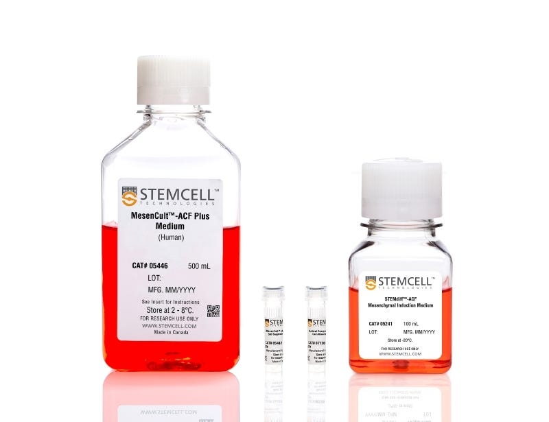
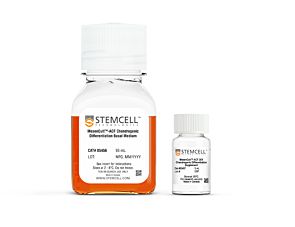
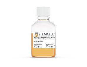
STEMdiff™ Mesenchymal Progenitor Kit
Defined culture kit for derivation and expansion of mesenchymal progenitor cells
Request Pricing
Thank you for your interest in this product. Please provide us with your contact information and your local representative will contact you with a customized quote. Where appropriate, they can also assist you with a(n):
Estimated delivery time for your area
Product sample or exclusive offer
In-lab demonstration
Overview
CollPlant is the manufacturer of the rhCollagen component of the cell attachment substrate.
Data Figures

Figure 1. Schematic of Differentiation Protocol and Timeline
In Phase 1, human ES or iPS cells are cultured in mTeSR™1 or TeSR™-E8™ medium. On Day 0 (Phase 2) of the protocol, cells are ready for induction into early mesoderm progenitor cells by replacing TeSR™ medium with STEMdiff™ Mesenchymal Induction Medium. By Day 4 (Phase 3), STEMdiff™ Mesenchymal Induction Medium is replaced with MesenCult™-ACF Medium to derive early mesenchymal progenitor cells (MPCs). On Day 6, cells are passaged onto cultureware precoated with MesenCult™-ACF Attachment Substrate in MesenCult™-ACF Medium. By Day 21, human ES- or iPS-derived MPCs exhibit the suggested MPC characteristics.

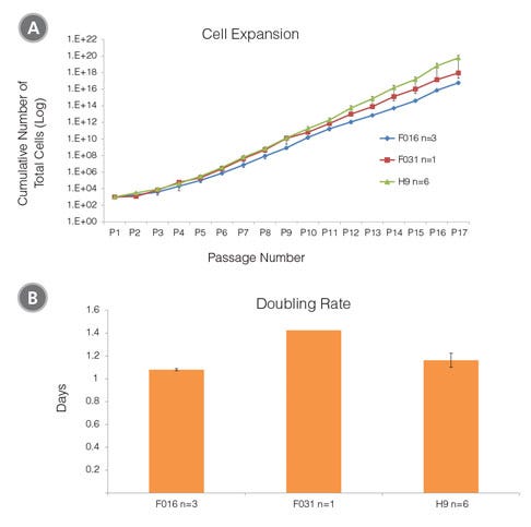
Figure 2. Cell Expansion and Doubling Rate of MPCs Derived from Human ES (H9) and iPS (STiPS-F016 and -F031) Cells in MesenCult™-ACF Medium
(A) The average cell expansion per passage over 17 passages for MPCs derived from human ES and iPS cell lines are approximately 9 and 10 fold. (B) Days to double cell number for human ES- and iPS-derived MPCs range from 1.1 to 1.4 days.

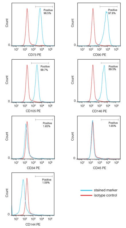
Figure 3. A Representative Flow Cytometric Analysis of STiPS-F016-derived MPCs Expressing Mesenchymal Surface Markers By Day 21
Human iPS-derived MPCs, generated using the STEMdiff™ Mesenchymal Progenitor Kit, express high levels of mesenchymal surface markers (CD73, CD90 and CD105) and the perivascular marker, CD146. MPCs do not express hematopoietic (CD34, CD45) and endothelial (CD144) surface markers. Human ES-derived MPCs express the same phenotype (data not shown).

Figure 4. Human ES- and iPS-derived MPCs Can Be Further Differentiated Into Adipogenic, Chondrogenic and Osteogenic Lineages
(A) MPCs generated from the 3 week protocol (described in Figure 1) and subsequently cultured in MesenCult™-ACF Medium develop MPC-like morphology (40X magnification). MPCs can be differentiated to (B) adipocytes (Oil Red O staining), 400X magnification; (C) chondrocytes (Alcian Blue staining), 100X magnification; and (D) osteoblasts (Fast Red and Silver Nitrate staining), 100X magnification.
Protocols and Documentation
Find supporting information and directions for use in the Product Information Sheet or explore additional protocols below.
Applications
This product is designed for use in the following research area(s) as part of the highlighted workflow stage(s). Explore these workflows to learn more about the other products we offer to support each research area.
Resources and Publications
Educational Materials (8)
Publications (6)
Abstract
Abstract
Abstract
Quality Statement:
PRODUCTS ARE FOR RESEARCH USE ONLY AND NOT INTENDED FOR HUMAN OR ANIMAL DIAGNOSTIC OR THERAPEUTIC USES UNLESS OTHERWISE STATED. FOR ADDITIONAL INFORMATION ON QUALITY AT STEMCELL, REFER TO WWW.STEMCELL.COM/COMPLIANCE.