mTeSR™1

cGMP, feeder-free maintenance medium for human ES and iPS cells

New look, same high quality and support! You may notice that your instrument or reagent packaging looks slightly different from images displayed on the website, or from previous orders. We are updating our look but rest assured, the products themselves and how you should use them have not changed. Learn more

mTeSR™1

cGMP, feeder-free maintenance medium for human ES and iPS cells

From: 364 USD
Catalog #
(Select a product)
cGMP, feeder-free maintenance medium for human ES and iPS cells
Add to Wish List

What's Included

  • mTeSR™1 Complete Kit (Catalog #85850)
    • mTeSR™1 Basal Medium, 400 mL
    • mTeSR™1 5X Supplement, 100 mL
  • mTeSR™1 Complete Kit, 1 L (Catalog #85857)
    • mTeSR™1 Basal Medium, 800 mL
    • mTeSR™1 5X Supplement, 100 mL, 2 Bottles

What Our Scientist Says

It makes me proud knowing that my work is critical to keeping thousands of hPSC lines reliably healthy and consistent around the world.

Arwen HunterAssociate Director, Stem Cell Biology
Arwen Hunter, Associate Director, Stem Cell Biology

Overview

Use this specialized, feeder-free culture medium to achieve more consistent human pluripotent stem cell (hPSC) cultures with homogenous, undifferentiated phenotypes.

Manufactured under relevant cGMPs, mTeSR™1 ensures the highest quality and consistency for reproducible results in your fundamental research, as well as for cell therapy and investigational new drug research applications. This serum-free, complete cell culture medium is made with pre-screened raw materials to ensure batch-to-batch consistency and robust performance in feeder-free hPSC culture.

Use established protocols for applications ranging from derivation to differentiation with this most widely published feeder-free hPSC culture medium, which has been used by leading pluripotent stem cell researchers to successfully maintain thousands of hPSC lines in over 50 countries. For enhanced cell performance and versatile maintenance, you may also be interested in mTeSR™ Plus medium, which is also manufactured under relevant cGMPs and features stabilized components and enhanced buffering.

To request a Letter of Authorization (LOA) for the FDA Master File for mTeSR™1, click here.
Subtype
Specialized Media
Cell Type
Pluripotent Stem Cells
Species
Human
Application
Cell Culture, Expansion, Maintenance
Brand
TeSR
Area of Interest
Stem Cell Biology
Formulation Category
Serum-Free

Data Figures

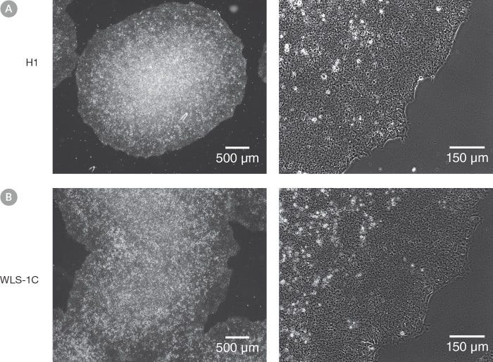
Figure 1. Normal hES and hiPS Cell Morphology is Observed in cGMP mTeSR™1 Cultures

Undifferentiated (A) H1 human embryonic stem (hES) and (B) WLS-1C human induced pluripotent stem (hiPS) cells cultured on Corning® Matrigel® Matrix in cGMP mTeSR™1 retain the prominent nucleoli and high nuclear-to-cytoplasmic ratio characteristic of this cell type after 10 passages. Densely packed cells and multi-layering are prominent when cells are ready to be passaged.

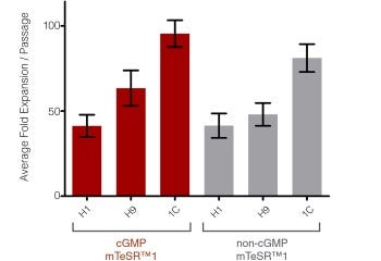
Figure 2. High Expansion Rates are Observed in cGMP mTeSR™1 Cultures

Graph shows the average fold expansion per passage +/- SEM obtained for hES (H1 and H9) and hiPS (WLS-1C) cells cultured in cGMP mTeSR­™1 (red) or non-cGMP mTeSR™1 (gray) on Corning® Matrigel® Matrix over 10 passages. Expansion was determined by enumerating the cell aggregates obtained at harvest and dividing by the number of cell aggregates seeded. Note that this data is representative of cultures passaged after 6-7 days in culture, lower expansion should be expected if using shorter culture times.

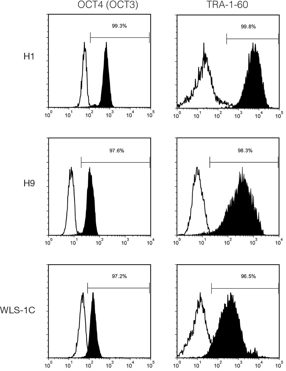
Figure 3. Cells Cultured in cGMP mTeSR™1 Medium Express Undifferentiated Cell Markers

Histogram analysis for hES (H1 and H9) and hiPS (WLS-1C) cells characterized using FACS for undifferentiated cell markers, OCT4 (OCT3) (Catalog #60093) and TRA-1-60 (Catalog #60064), after 8 - 10 passages in cGMP mTeSR™1 (filled = sample, blank = isotype control).

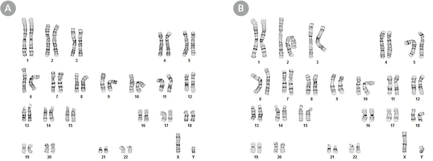
Figure 4. hPSCs Maintained in cGMP mTeSR™1 Display a Normal Karyotype

Karyograms of (A) H1 hES and (B) WLS-1C hiPS cells cultured in cGMP mTeSR™1 for 11 passages shows that a normal karyotype is retained.

Protocols and Documentation

Find supporting information and directions for use in the Product Information Sheet or explore additional protocols below.

Document Type
Product Name
Catalog #
Lot #
Language
Product Name
mTeSR™1
Catalog #
85857, 85850
Lot #
All
Language
English
Document Type
Technical Manual
Product Name
mTeSR™1
Catalog #
85850
Lot #
All
Language
English
Document Type
Safety Data Sheet 1
Product Name
mTeSR™1
Catalog #
85857, 85850
Lot #
All
Language
English
Document Type
Safety Data Sheet 2
Product Name
mTeSR™1
Catalog #
85857, 85850
Lot #
All
Language
English
Document Type
Safety Data Sheet 3
Product Name
mTeSR™1
Catalog #
85857, 85850
Lot #
All
Language
English
The Certificate of Analysis for this product has been updated for newly released materials. To access respective CoAs please use this tool.

Applications

This product is designed for use in the following research area(s) as part of the highlighted workflow stage(s). Explore these workflows to learn more about the other products we offer to support each research area.

Resources and Publications

Educational Materials (41)

Publications (1583)

iPSC-Based Modeling of RAG2 Severe Combined Immunodeficiency Reveals Multiple T Cell Developmental Arrests. M. Themeli et al. Stem cell reports 2020 feb

Abstract

RAG2 severe combined immune deficiency (RAG2-SCID) is a lethal disorder caused by the absence of functional T and B cells due to a differentiation block. Here, we generated induced pluripotent stem cells (iPSCs) from a RAG2-SCID patient to study the nature of the T cell developmental blockade. We observed a strongly reduced capacity to differentiate at every investigated stage of T cell development, from early CD7-CD5- to CD4+CD8+. The impaired differentiation was accompanied by an increase in CD7-CD56+CD33+ natural killer (NK) cell-like cells. T cell receptor D rearrangements were completely absent in RAG2SCID cells, whereas the rare T cell receptor B rearrangements were likely the result of illegitimate rearrangements. Repair of RAG2 restored the capacity to induce T cell receptor rearrangements, normalized T cell development, and corrected the NK cell-like phenotype. In conclusion, we succeeded in generating an iPSC-based RAG2-SCID model, which enabled the identification of previously unrecognized disorder-related T cell developmental roadblocks.
IGF1-mediated human embryonic stem cell self-renewal recapitulates the embryonic niche. S. E. Wamaitha et al. Nature communications 2020

Abstract

Our understanding of the signalling pathways regulating early human development is limited, despite their fundamental biological importance. Here, we mine transcriptomics datasets to investigate signalling in the human embryo and identify expression for the insulin and insulin growth factor 1 (IGF1) receptors, along with IGF1 ligand. Consequently, we generate a minimal chemically-defined culture medium in which IGF1 together with Activin maintain self-renewal in the absence of fibroblast growth factor (FGF) signalling. Under these conditions, we derive several pluripotent stem cell lines that express pluripotency-associated genes, retain high viability and a normal karyotype, and can be genetically modified or differentiated into multiple cell lineages. We also identify active phosphoinositide 3-kinase (PI3K)/AKT/mTOR signalling in early human embryos, and in both primed and na{\{i}}ve pluripotent culture conditions. This demonstrates that signalling insights from human blastocysts can be used to define culture conditions that more closely recapitulate the embryonic niche."
Maturation of Human Pluripotent Stem Cell-Derived Cerebellar Neurons in the Absence of Co-culture. T. P. Silva et al. Frontiers in bioengineering and biotechnology 2020

Abstract

The cerebellum plays a critical role in all vertebrates, and many neurological disorders are associated with cerebellum dysfunction. A major limitation in cerebellar research has been the lack of adequate disease models. As an alternative to animal models, cerebellar neurons differentiated from pluripotent stem cells have been used. However, previous studies only produced limited amounts of Purkinje cells. Moreover, in vitro generation of Purkinje cells required co-culture systems, which may introduce unknown components to the system. Here we describe a novel differentiation strategy that uses defined medium to generate Purkinje cells, granule cells, interneurons, and deep cerebellar nuclei projection neurons, that self-formed and differentiated into electrically active cells. Using a defined basal medium optimized for neuronal cell culture, we successfully promoted the differentiation of cerebellar precursors without the need for co-culturing. We anticipate that our findings may help developing better models for the study of cerebellar dysfunctions, while providing an advance toward the development of autologous replacement strategies for treating cerebellar degenerative diseases.
New look, same high quality and support! You may notice that your instrument or reagent packaging looks slightly different from images displayed on the website, or from previous orders. We are updating our look but rest assured, the products themselves and how you should use them have not changed. Learn more