IntestiCult™ Organoid Growth Medium (Human)

Cell culture medium for establishment and maintenance of human intestinal organoids
Interested in trying STEMCELL’s organoid products for your intestinal research? Fill out the form to request information about introductory offers.

IntestiCult™ Organoid Growth Medium (Human)

Cell culture medium for establishment and maintenance of human intestinal organoids

From: 550 USD
Catalog #
(Select a product)
Cell culture medium for establishment and maintenance of human intestinal organoids
Add to Wish List

Product Advantages


  • Convenient, in vitro system that recapitulates many key characteristics of the adult intestinal epithelium, including intra- and intercellular signaling, self-propagating stem cell niche, and functional transport into and out of the lumen

  • Complete medium formulation that delivers consistent results

  • Enables generation of intestinal organoids in one week

  • Easy-to-use format and optimized protocol

What's Included

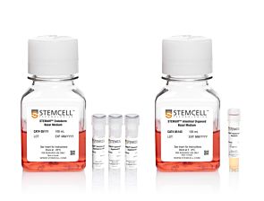
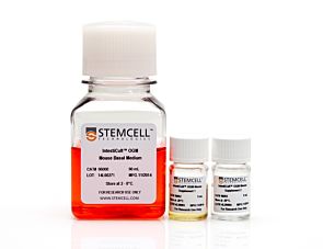
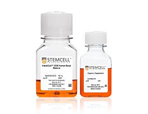
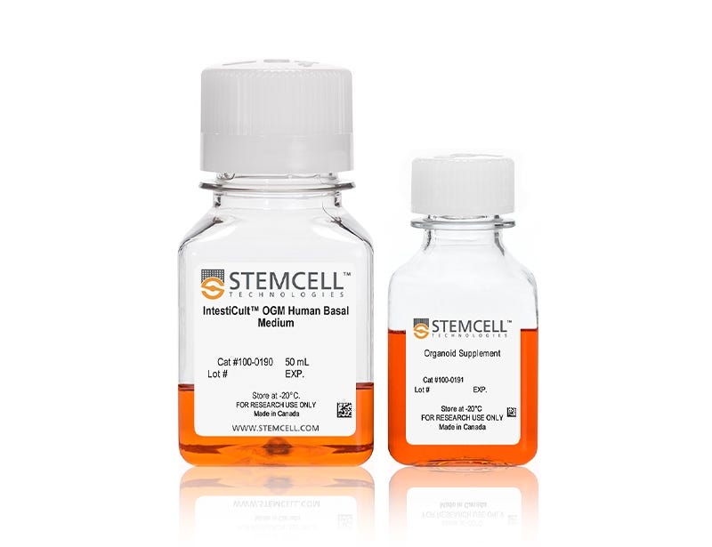
  • IntestiCult™ OGM Human Basal Medium, 50 mL
  • Organoid Supplement, 50 mL
Products for Your Protocol
To see all required products for your protocol, please consult the Protocols and Documentation.

What Our Scientist Says

Organoids have truly expanded the limits of what's possible for in vitro studies of the intestinal epithelium. By providing optimized culture media and robust, approachable protocols, we are making these technologies more accessible to researchers.

Ryan ConderAssociate Director, Gastrointestinal Biology
Ryan Conder, Associate Director

Overview

Model key characteristics of the adult intestinal epithelium using intestinal organoids established and maintained with this complete medium formulation and optimized protocol. Using the easy-to-follow and robust protocol, you can derive organoids from human intestinal crypts in one week; organoid growth across donor samples, including those that are otherwise difficult to grow, is enabled by an enriched stem cell population.

Organoids grown in IntestiCult™ Organoid Growth Medium (Human) incorporate a functional lumen enclosed by a polarized intestinal epithelial cell layer and, for versatile modeling applications, can be further differentiated in 3D or in 2D as submerged monolayers or air-liquid interface (ALI) cultures using IntestiCult™ Organoid Differentiation Medium (Human; Catalog #100-0214).

Applications of intestinal organoid cultures include studying the development and function of intestinal epithelium, modeling intestinal diseases, and screening molecules for both efficacy and toxicity in an intestinal model. Intestinal organoid cultures can also be used for investigation of adult stem cell properties and for regenerative therapy approaches.

Learn how to culture human intestinal organoids in our On-Demand Intestinal Course or browse our Frequently Asked Questions (FAQs) about the organoid workflows using IntestiCult™. Additionally, download our detailed e-book Proven Protocols for Intestinal Organoid Culture: Getting Started with IntestiCult™ for a curated collection of intestinal organoid protocols.

Should you intend to use this product for commercial purposes, please contact HUB Organoids B.V. at www.huborganoids.nl for a commercial use license or for clarifications in relation to HUB Organoids B.V. licensing.
Subtype
Specialized Media
Cell Type
Intestinal Cells
Species
Human
Application
Cell Culture, Differentiation, Expansion, Maintenance, Organoid Culture
Brand
IntestiCult
Area of Interest
Disease Modeling, Epithelial Cell Biology, Stem Cell Biology

Data Figures

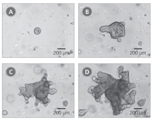
Figure 1. Primary Organoids Grown in IntestiCult™ Organoid Growth Medium (Human) are Fully Mature After 10-14 Days in Culture

Primary organoids were cultured from human colonic biopsy samples and grown in IntestiCult Organoid Growth Medium (Human). Organoids were imaged after (A) two days, (B) six days, (C) eight days and (D) ten days growth.

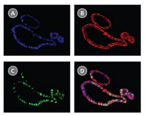
Figure 2. Organoids Grown in IntestiCult™ Organoid Growth Medium (Human) Display Markers of Human Intestinal Epithelial Cells

Immunofluorescence of organoids grown in IntestiCult™ Organoid Growth Medium (Human) showing colocalization of (A) DAPI, (B) EPCAM and (C) Ki67. (D) A merged image shows the position of actively proliferating (Ki67+) intestinal stem cells within the epithelial layer (EPCAM+).

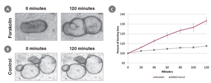
Figure 3. Forskolin-Induced Swelling of Organoids Grown in IntestiCult™ Organoid Growth Medium (Human)

Organoids were treated with (A) 5 μM Forskolin or (B) DMSO and organoid area was measured at 0 minutes and 120 minutes. (C)Forskolin-treated organoids increased in size 33.5 ± 3.8% compared to 7.5 ± 0.8% for DMSO-treated organoids.

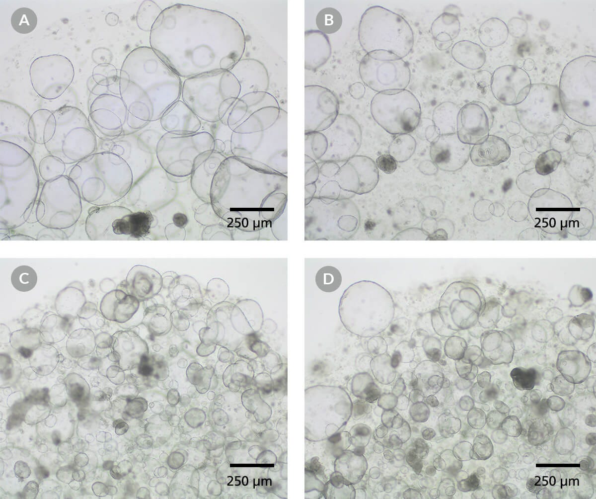
Figure 4. IntestiCult™ Organoid Growth Medium (Human) Supports the Growth of Organoids in Multiple Extracellular Matrices

Intestinal organoid cultures were prepared in IntestiCult™ Organoid Growth Medium (Human) and plated in (A) Matrigel® Growth Factor Reduced Basement Membrane Matrix (Corning® catalog # 356231), (B) Geltrex® LDEV-Free Reduced Growth Factor Basement Membrane Matrix (Gibco™ catalog # A1413202), (C) Cultrex® Reduced Growth Factor Basement Membrane Extract, Type 1 (R&D Systems™ catalog # 3433-005-R1), and (D) Cultrex® Reduced Growth Factor Basement Membrane Extract, Type 2 (R&D Systems™ catalog # 3533-005-02). Organoid cultures are imaged at the end of passage 4. All four extracellular matrices supported robust growth of human intestinal organoids. Scale bars = 250 μm.

Data and images of the MIMETAS OrganoReady Colon Organoid Plate

Figure 5. The MIMETAS OrganoReady® Colon Organoid Platform Uses IntestiCult™ to Create an Advanced Physiologically Relevant Model for Gastrointestinal Toxicity Testing and Barrier Integrity

(A) The OrganoReady® plate highlighting the microfluidic compartments.

(B) Schematic of the OrganoReady® microfluidic compartments where columns 1, 2, and 3 house the medium, a collagen-1 matrix, and the colon organoid tubule, respectively.

(C) Immunofluorescence staining of the colon organoid tubule confirms an adult tissue phenotype with the presence of goblet cells (Muc2), enterocytes (Occludin), and stem cells (Sox9). The 3D-lumenized structure provides apical (Ezrin) and basolateral (Integrin-β4) access to the polarized epithelium. Additionally, the organoid tubules show polarized and modulatable activity of expression of P-glycoprotein (Pgp).

(D) The OrganoReady® Colon Organoid platform supports toxicity testing, as demonstrated by dose-dependent measurements of TEER, LDH, and ATP following exposure to Afatinib (n = 4, N = 2). After 72 hrs of exposure, a dose dependent decrease in TEER, cytotoxicity, and cell viability was observed. For more information, please visit mimetas.com/en/organoready-organoid/.

Protocols and Documentation

Find supporting information and directions for use in the Product Information Sheet or explore additional protocols below.

Document Type
Product Name
Catalog #
Lot #
Language
Catalog #
06010
Lot #
All
Language
English
Document Type
Safety Data Sheet 1
Catalog #
06010
Lot #
All
Language
English
Document Type
Safety Data Sheet 2
Catalog #
06010
Lot #
All
Language
English
Document Type
Safety Data Sheet 3
Catalog #
06010
Lot #
All
Language
English
Document Type
Safety Data Sheet 4
Catalog #
06010
Lot #
All
Language
English

Applications

This product is designed for use in the following research area(s) as part of the highlighted workflow stage(s). Explore these workflows to learn more about the other products we offer to support each research area.

Resources and Publications

Educational Materials (62)

Publications (49)

An integrated patient-derived colon organoids platform as a functional model for nutraceutical and stress response. A. Costantino et al. iScience 2026 Jun

Abstract

Nutraceuticals are increasingly investigated for their capacity to modulate oxidative and inflammatory stress, yet preclinical testing still relies largely on immortalized cell lines or animal models that poorly recapitulate human epithelial complexity. To address this gap, we developed an integrated platform based on patient-derived colon organoids generated from non-tumoral mucosa and maintained under proliferative or differentiation conditions to model distinct epithelial states. The system combines millifluidic measurement of individual organoid mass, density, and diameter with bulk RNA sequencing and digital PCR profiling to enable multiparametric characterization. Transcriptional analysis revealed state-specific gene programs and shifts in epithelial and immune-related pathways, while biophysical measurements captured structural remodeling. In this pilot validation, a defined oxidative insult followed by nutraceutical treatment elicited coordinated transcriptional and phenotypic responses. This integrated approach provides a scalable and physiologically relevant framework for functional nutraceutical profiling and mechanistic studies of epithelial stress responses.
12-HHT is associated with epithelial barrier enhancement and reduced inflammatory responses in colon organoids of normoganglionosis in Hirschsprung's disease. K. Suda et al. PloS one 2026 Jul

Abstract

PURPOSE: Hirschsprung-associated enterocolitis remains a major postoperative complication of Hirschsprung's disease (HD), and impaired epithelial barrier integrity has been proposed as a contributing factor. In this study, we investigated whether 12-hydroxyheptadecatrienoic acid (12-HHT), an endogenous leukotriene B4 receptor 2 (BLT2) agonist, is associated with epithelial barrier enhancement and reduced inflammatory responses in patient-derived colonic organoids. METHODS: Normoganglionic specimens from rectal/rectosigmoid HD at pull-through (HD-N; n = 8) and transverse colon specimens from anorectal malformation (ARM) at colostomy closure (n = 10) were used to generate colonic organoids. Epithelia were isolated using ethylenediaminetetraacetic acid and subsequently embedded in Matrigel. Baseline expression of TJP1, TJP2, F11R (encoding junctional adhesion molecule-A), JAM2, CLDN1, CLDN3, CLDN4) and LTB4R2 (encoding BLT2) was assessed by qPCR and immunoblotting. Organoids were then treated with 12-HHT (0.4, 2, or 10 μM) for 7 days, followed by qPCR. Additional experiments assessed cytokine expression (IL1B, IL6) and TJPs after 24 h with tumor necrosis factor-α (TNF-α, 100 ng/mL) plus phosphate buffered saline or 12-HHT. Barrier function was evaluated using FITC-dextran influx assays. RESULTS: HD-N and ARM organoids exhibited similar growth efficiencies. Baseline expression for F11R, JAM2, CLDN1, CLDN3, CLDN4, and LTB4R2 was significantly lower in HD-N than in ARM. TJPs were upregulated by 12-HHT at 2 and 10 μM in both groups, with stronger effects in ARM. In HD-N organoids, 10 μM 12-HHT suppressed TNF-α-induced IL1B and IL6 elevation mitigated tight junction proteins (TJPs) downregulation more effectively than 2 μM. 12-HHT attenuated TNF-α-induced FITC-dextran influx in HD-N organoids. CONCLUSION: 12-HHT is associated with epithelial barrier enhancement and reduced inflammatory responses in HD-N organoids.
A Colon Cancer Organoid-on-a-Chip Model for In Vitro Therapy Assessment. L. Valle et al. International journal of molecular sciences 2026 Jul

Abstract

Colon cancer is one of the leading causes of death, requiring advanced therapies that need models for developing new drugs. Conventional cell culture models do not accurately and precisely reproduce the complexity of the tumor microenvironment, limiting their usefulness in research and therapy development. To address this weakness, patient-derived organoids have emerged as promising in vitro models. The implementation of these organoid-based models into more physiologically relevant systems is expected to improve their clinical relevance. Thus, integrating these organoids into microfluidic chips acting as bioreactors will likely improve the predictive response of therapies in personalized medicine. In this article, we report the development of a new colon cancer organoid-on-a-chip model that enables the in vitro culture of patient-derived colon cancer organoids under continuous culture media flow. We demonstrate how the developed organoids were derived from tumor biopsies of patients with colorectal cancer, expanded in standard three-dimensional (3D) culture, and cultured inside the microfluidic chips. The microfluidic chip chambers are designed to house organoids in a controlled environment, allowing the injection of therapies and monitoring by optical microscopy in real time. The in vitro therapies tested were a combination of drugs based on 5-fluorouracil and oxaliplatin at different concentrations. As a result, we demonstrate for the first time that this model proves the capability of this technology for in vitro testing colon cancer therapies.
Interested in trying STEMCELL’s organoid products for your intestinal research? Fill out the form to request information about introductory offers.