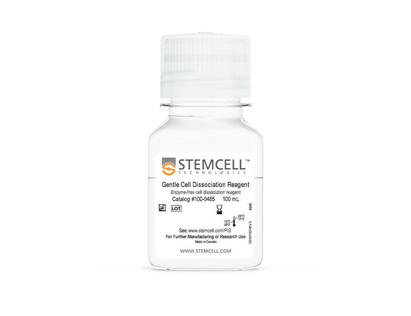
Gentle Cell Dissociation Reagent

cGMP, enzyme-free cell dissociation reagent

Gentle Cell Dissociation Reagent

cGMP, enzyme-free cell dissociation reagent
From: 62 USD
Catalog #
(Select a product)
cGMP, enzyme-free cell dissociation reagent
Add to Wish List

Product Advantages


  • Obtain quality cultures with this gentle, chemically-defined, enzyme-free, GMP solution

  • Achieve high expansion of human ES/iPS cells during routine culture

  • Simplify cell passaging with straightforward, room temperature passaging protocols

Overview

Use Gentle Cell Dissociation Reagent (GCDR) to dissociate human embryonic stem (ES) cells or human induced pluripotent stem (iPS) cells into cell aggregates for routine passaging, or into a single-cell suspension. This enzyme-free and chemically-defined reagent ensures high expansion of human ES and iPS cells during routine culture. It is also suitable for the isolation of intestinal crypts to establish intestinal organoids, and for breaking up Corning® Matrigel® domes when passaging organoid cultures.

GCDR is now also manufactured following relevant cGMPs under a certified quality management system to ensure the highest quality and consistency for reproducible results.

To request a Letter of Authorization (LOA) for Gentle Cell Dissociation Reagent’s Drug Master File, click here.
Subtype
Non-Enzymatic
Cell Type
Endoderm, PSC-Derived, Intestinal Cells, Pluripotent Stem Cells
Species
Human, Mouse
Application
Cell Culture
Area of Interest
Epithelial Cell Biology, Stem Cell Biology

Protocols and Documentation

Find supporting information and directions for use in the Product Information Sheet or explore additional protocols below.

Document Type
Product Name
Catalog #
Lot #
Language
Catalog #
100-0485, 100-1077
Lot #
Lot 1000157162 and higher For 100-0485 | Lot 1000157164 and higher For 100-1077
Language
English
Catalog #
100-0485, 100-1077
Lot #
Lot 1000157161 and lower For 100-0485 | Lot 1000157163 and lower For 100-1077
Language
English
Document Type
Safety Data Sheet
Catalog #
100-0485
Lot #
All
Language
English
Document Type
Safety Data Sheet
Catalog #
100-1077
Lot #
All
Language
English

Resources and Publications

Publications (120)

Gliomas phenocopy an inborn error of metabolism to drive neuronal activity and tumor growth. K. Abdullah et al. Cell 2026 Sep

Abstract

The metabolic hallmarks of high-grade glioma (HGG) are not fully understood. Human brain tissue metabolomics revealed that the creatine synthesis pathway intermediate guanidinoacetate (GAA) accumulated ∼100-fold in HGGs relative to controls, which was caused by imbalanced activities of enzymes in this pathway. Glioma cells secreted GAA rather than using it to produce creatine, implicating an alternative function. GAA accumulates in GAA N-methyltransferase (GAMT) deficiency, an inborn error of metabolism, and elevates neuronal excitability. Neuronal excitability is also increased in glioma and drives tumor growth through neuron-glioma interactions. We hypothesized that glioma-generated GAA excites surrounding neurons. Indeed, GAA induced neuronal hyperactivity by activating GABAA receptors and causing depolarizing currents in glioma-associated neurons with dysregulated chloride homeostasis. Depleting tumoral GAA decreased electrochemical activity, neuron-glioma interactions, and tumor aggressiveness. Our findings unveil a mechanism linking cancer metabolism with cancer neuroscience and leverage human genetics to nominate GAA synthesis as a target in gliomas.
ALY688 Protects Against Myocardial Ischemia-Reperfusion Injury via Direct Effects and Rab8a-Dependent Extracellular Vesicles. H. Sung et al. Journal of extracellular vesicles 2026 Sep

Abstract

Despite advances in percutaneous coronary intervention, ischemia-reperfusion (IR) injury remains a major cause of morbidity and mortality. Adiponectin confers broad cardioprotective effects, motivating the development of adiponectin receptor agonists. Here, we investigated the cardioprotective efficacy and mechanisms of ALY688, a synthetic adiponectin receptor agonist peptide, in myocardial IR injury. In a clinically translatable rat IR model, intravenous administration of ALY688 during ischemia together with subcutaneous dosing that continued for 28 days reduced troponin-I levels, cardiomyocyte death, and infarct size, while preserving cardiac function. ALY688 restored autophagic flux, mitigated reactive oxygen species accumulation, and suppressed apoptosis in both IR hearts and hypoxia-reoxygenation (HR)-treated cardiomyocytes. Proteomic profiling revealed that Rab8a, downregulated by IR, was maintained with ALY688 treatment. Notably, ALY688 increased extracellular vesicle (EV) abundance in myocardium and plasma, and EVs from treated animals displayed distinct proteomic signatures enriched in glycolytic and oxidative stress-related proteins. These EVs conferred protection against HR-induced injury in H9c2 and human iPSC-derived cardiomyocytes. CRISPR-mediated Rab8a knockout impaired ALY688-induced EV biogenesis and attenuated the cytoprotective effects of these EVs. Collectively, these findings identify ALY688 as a promising therapeutic that mitigates IR injury via both direct myocardial protection and Rab8a-dependent EV-mediated cardioprotective signalling.
NheABC is a pH-dependent cytotoxin that targets mitochondria and contributes to the virulence of Bacillus cereus in wound infections. N. Ullah et al. Cell communication and signaling : CCS 2026 Sep

Abstract

Members of the Bacillus cereus group are Gram-positive, spore-forming, facultative anaerobic bacteria widely distributed in the environment. Pathogenic B. cereus group strains have been linked to a range of illnesses and infections, including severe non-gastrointestinal infections, such as wound infections. In the present study, we show that, among the pore-forming toxin (PFT)-producing bacterial strains tested, B. cereus group strain 75/95 induced pronounced membrane permeabilization in epithelial cells, prompting us to investigate the contribution of its tripartite NheABC toxin to host cell damage. NheABC exhibited potent cell membrane permeability in 2D epithelial cell cultures as well as in 3D spheroids and intestinal organoids, with prominent effects on the cell membrane and mitochondria. Functional analysis using liposome leakage assays demonstrated that cardiolipin (CL) and phosphatidylethanolamine (PE) promote NheABC-mediated membrane permeabilization, suggesting that membrane lipid composition contributes to NheABC activity and mitochondrial damage. Moreover, using erythrocytes and epithelial cells as model systems, we further demonstrated that the cytolytic activity of NheABC is pH-dependent and is markedly reduced under acidic conditions (pH 5.5). This pH dependence was independently confirmed using liposome leakage assays. Importantly, NheABC enhanced bacterial colonization and the local inflammatory response in a murine model of wound infection. Overall, our study highlights the critical role of mitochondria and pH in regulating NheABC-mediated cytolytic activity in mammalian cells, which may lead to the development of novel therapeutic strategies for managing B. cereus group-related gastrointestinal and non-gastrointestinal infections.