Make more informed purchasing decisions with our new product availability and delivery estimate feature, now available on all product pages, in your cart, and during checkout.
Sign In
New to STEMCELL?
Register for an account to quickly and easily purchase products online and for one-click access to all educational content.
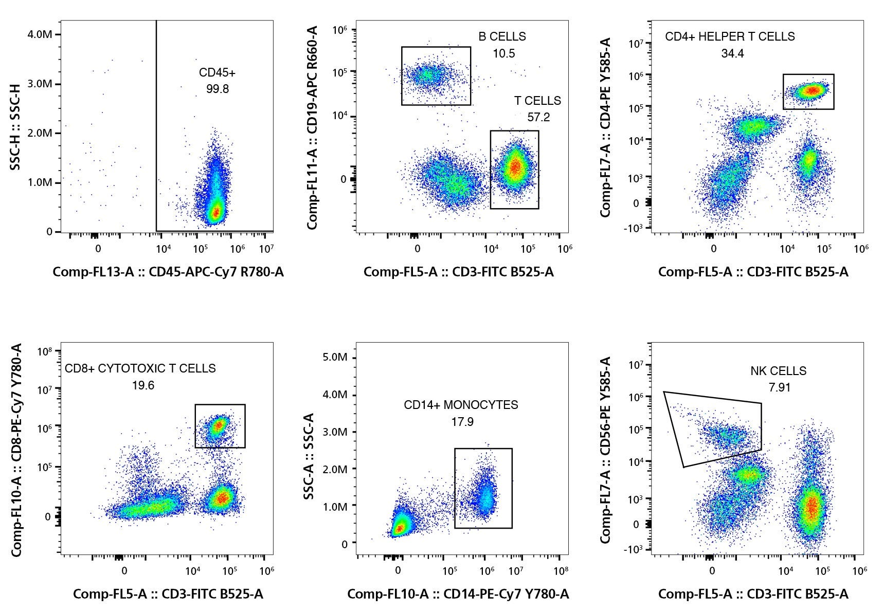
Maximize your savings on high-quality PBMCs! Take advantage of our bulk pricing discounts and price matching offer to get the best value for your purchase. Now available for specific lots, the standard flow cytometry panel provides the exact frequencies of common cell types in each vial of PBMCs. See Figure 1 in the Data Figures section below to view a representative panel. Browse our Frequently Asked Questions for more information.
Request Pricing
Thank you for your interest in this product.
Please provide us with your contact information and your local representative
will contact you with a customized quote. Where appropriate, they can also assist you with a(n):
Estimated delivery time for your area
Product sample or exclusive offer
In-lab demonstration
By submitting this form, you are providing your consent to STEMCELL Technologies Canada Inc. and its subsidiaries and affiliates (“STEMCELL”) to collect and use your information, and send you newsletters and emails in accordance with our privacy policy. Please contact us with any questions that you may have. You can unsubscribe or change your email preferences at any time.
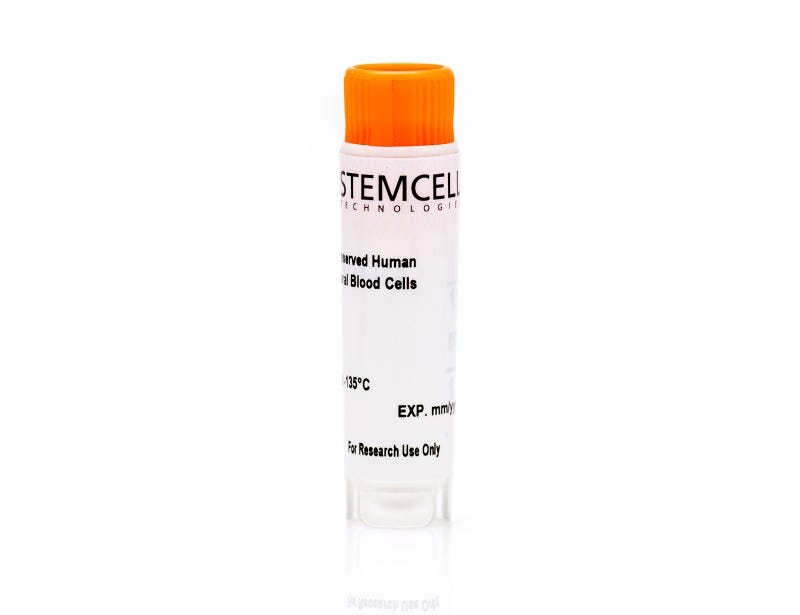
Streamline your assays with ready-to-use, ethically sourced, primary human mononuclear cells. With personalized service, custom products, flexible delivery times, and the option to reserve entire lots to prescreen cells for applications, we help you get the cells you need.
Isolated from peripheral blood leukapheresis samples using density gradient separation and/or red blood cell lysis and cryopreserved in animal component-free CryoStor®CS10 medium (Catalog #07930), cells are collected using Institutional Review Board (IRB)- or Research Ethics Committee (REC)-approved consent forms and protocols. Additional documentation and high-resolution HLA typing (Class I and Class II alleles and CMV status) are available upon request. Acid-citrate-dextrose solution A (ACDA) is added during collection as an anticoagulant. Donor specifications (e.g. BMI category, smoking status, ethnicity, etc.) can be requested in the comment box above, after selecting from the product options. Donors are screened for HIV-1, HIV-2, hepatitis B, and hepatitis C.
Certain products are only available in select territories. Please contact your local sales representative or Product & Scientific Support at techsupport@stemcell.com for further information.
Figure 1. Typical Flow Cytometric Analysis Profile of PBMCs
Representative gating strategy of immune cell populations present in PBMCs. Flow cytometry was performed on the peripheral blood mononuclear cells (PBMCs) post-thaw and can be provided for specific lots. The CD45 plot was gated on viable single cells while all other plots were gated on viable CD45+ single cells. In the above example, the cell frequencies are as follows: leukocytes (CD45+), 99.8%; B cells (CD19+), 10.5%; T Cells (CD3+), 57.2%; helper T cells (CD3+CD4+), 34.4%; Cytotoxic T cells (CD3+CD8+), 19.6%; monocytes (CD14+), 17.9%; and NK cells (CD3-CD56+), 7.91%.
This product is designed for use in the following research area(s) as part
of the highlighted workflow stage(s). Explore these workflows to learn more about the other products we
offer to support each research area.
Eradication of Triple-Negative Breast Cancer Cells by Targeting Glycosylated PD-L1.
C.-W. Li et al.
Cancer cell 2018 FEB
Abstract
Protein glycosylation provides proteomic diversity in regulating protein localization, stability, and activity; it remains largely unknown whether the sugar moiety contributes to immunosuppression. In the study of immune receptor glycosylation, we showed that EGF induces programmed death ligand 1 (PD-L1) and receptor programmed cell death protein 1 (PD-1) interaction, requiring beta$-1,3-N-acetylglucosaminyl transferase (B3GNT3) expression in triple-negative breast cancer. Downregulation of B3GNT3 enhances cytotoxic T cell-mediated anti-tumor immunity. A monoclonal antibody targeting glycosylated PD-L1 (gPD-L1) blocks PD-L1/PD-1 interaction and promotes PD-L1 internalization and degradation. In addition to immune reactivation, drug-conjugated gPD-L1 antibody induces a potent cell-killing effect as well as a bystander-killing effect on adjacent cancer cells lacking PD-L1 expression without any detectable toxicity. Our work suggests targeting protein glycosylation as a potential strategy to enhance immune checkpoint therapy.
Dendritic Cells but Not Macrophages Sense Tumor Mitochondrial DNA for Cross-priming through Signal Regulatory Protein α Signaling.
Xu MM et al.
Immunity 2017 AUG
Abstract
Inhibition of cytosolic DNA sensing represents a strategy that tumor cells use for immune evasion, but the underlying mechanisms are unclear. Here we have shown that CD47-signal regulatory protein α (SIRPα) axis dictates the fate of ingested DNA in DCs for immune evasion. Although macrophages were more potent in uptaking tumor DNA, increase of DNA sensing by blocking the interaction of SIRPα with CD47 preferentially occurred in dendritic cells (DCs) but not in macrophages. Mechanistically, CD47 blockade enabled the activation of NADPH oxidase NOX2 in DCs, which in turn inhibited phagosomal acidification and reduced the degradation of tumor mitochondrial DNA (mtDNA) in DCs. mtDNA was recognized by cyclic-GMP-AMP synthase (cGAS) in the DC cytosol, contributing to type I interferon (IFN) production and antitumor adaptive immunity. Thus, our findings have demonstrated how tumor cells inhibit innate sensing in DCs and suggested that the CD47-SIRPα axis is critical for DC-driven antitumor immunity.
Phosphorylation of NEUROG3 Links Endocrine Differentiation to the Cell Cycle in Pancreatic Progenitors.
Krentz NAJ et al.
Developmental cell 2017 APR
Abstract
During pancreatic development, proliferating pancreatic progenitors activate the proendocrine transcription factor neurogenin 3 (NEUROG3), exit the cell cycle, and differentiate into islet cells. The mechanisms that direct robust NEUROG3 expression within a subset of progenitor cells control the size of the endocrine population. Here we demonstrate that NEUROG3 is phosphorylated within the nucleus on serine 183, which catalyzes its hyperphosphorylation and proteosomal degradation. During progression through the progenitor cell cycle, NEUROG3 phosphorylation is driven by the actions of cyclin-dependent kinases 2 and 4/6 at G1/S cell-cycle checkpoint. Using models of mouse and human pancreas development, we show that lengthening of the G1 phase of the pancreatic progenitor cell cycle is essential for proper induction of NEUROG3 and initiation of endocrine cell differentiation. In sum, these studies demonstrate that progenitor cell-cycle G1 lengthening, through its actions on stabilization of NEUROG3, is an essential variable in normal endocrine cell genesis.
Maximize your savings on high-quality PBMCs! Take advantage of our bulk pricing discounts and price matching offer to get the best value for your purchase. Now available for specific lots, the standard flow cytometry panel provides the exact frequencies of common cell types in each vial of PBMCs. See Figure 1 in the Data Figures section below to view a representative panel. Browse our Frequently Asked Questions for more information.
Quality Statement:
PRODUCTS ARE FOR RESEARCH USE ONLY AND NOT INTENDED FOR HUMAN OR ANIMAL DIAGNOSTIC OR THERAPEUTIC USES UNLESS OTHERWISE STATED.