Anti-Beta-Tubulin III Antibody, Clone AA10

Mouse monoclonal IgG2a antibody against human, mouse, rat beta­tubulin III

Anti-Beta-Tubulin III Antibody, Clone AA10

Mouse monoclonal IgG2a antibody against human, mouse, rat beta­tubulin III

From: 153 USD
Catalog #
(Select a product)
Mouse monoclonal IgG2a antibody against human, mouse, rat beta­tubulin III
Add to Wish List

Overview

The AA10 antibody reacts with beta-tubulin III, an ~50 - 55 kDa structural protein that is a constituent of tubulin. Tubulin is the major component of microtubules within the cytoskeleton and is assembled from heterodimers of alpha and beta tubulin subunits. The beta III isoform of tubulin, also known as neuron-specific class III beta-tubulin, is expressed primarily in neurons and is widely used as a marker to distinguish neurons from other cell types. Beta-tubulin III contributes to microtubule formation in neuronal cell bodies and axons, a function involving GTP binding, and plays roles in axonal transport, neuronal cell proliferation, and differentiation. It is highly expressed in several types of cancer and is a predictive and prognostic marker for various tumors, for example, being found in neoplastic but not in normal glial cells. The AA10 antibody is expected to recognize all mammalian homologs of beta-tubulin III.
Subtype
Primary Antibodies
Target Antigen
Beta-Tubulin III
Alternative Names
Class 3 beta-tubulin, class III beta-tubulin, MC1R, neuron-specific class 3 beta-tubulin, neuron-specific class III beta-tubulin, TUBB 3, TUBB3, tubulin beta 3, tubulin beta 4, tubulin beta III
Reactive Species
Human, Mouse, Rat
Conjugation
Alexa Fluor 488, Unconjugated
Host Species
Mouse
Cell Type
Neural Cells, PSC-Derived, Neurons
Species
Human, Mouse, Rat
Application
Flow Cytometry, Immunocytochemistry, Immunofluorescence, Immunohistochemistry, Immunoprecipitation, Western Blotting
Area of Interest
Neuroscience
Clone
AA10
Gene ID
10381
Isotype
IgG2a, kappa

Data Figures

Data for Unconjugated

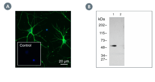
Figure 1. Data for Unconjugated

(A) E18 cortical rat neurons were cultured using the NeuroCult™ SM1 Neuronal Culture Kit on poly-lysine-coated glass coverslips, then fixed and labeled with Anti-Beta-Tubulin III Antibody, Clone AA10, followed by donkey anti-mouse IgG, Alexa Fluor® 488, and counter-stained with DAPI. Inset shows cells labeled with Mouse IgG2a, kappa Isotype Control Antibody, Clone MOPC-173 (Catalog #60071), followed by donkey anti-mouse IgG, Alexa Fluor® 488, and counter-stained with DAPI. (B) Western blot analysis of denatured/reduced cell lysates from adult rat brain cortical cells (lane 1) or mouse E13.5 neural progenitor cells cultured with NeuroCult™ Proliferation Kit (Mouse) (negative control, lane 2) with Anti-Beta-Tubulin III Antibody, Clone AA10.

Data for Alexa Fluor® 488-Conjugated

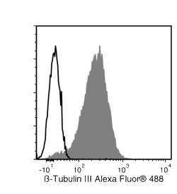
Figure 2. Data for Alexa Fluor® 488-Conjugated

E18 cortical rat neurons were cultured using the NeuroCult™ SM1 Neuronal Culture Kit on poly-lysine-coated glass coverslips, then fixed and labeled with Anti-Beta-Tubulin III Antibody, Clone AA10, Alexa Fluor® 488 (green), and counterstained with DAPI (blue). Inset shows cells incubated with a mouse IgG2a, kappa Alexa Fluor® 488 isotype control antibody, and counterstained with DAPI.

Protocols and Documentation

Find supporting information and directions for use in the Product Information Sheet or explore additional protocols below.

Document Type
Product Name
Catalog #
Lot #
Language
Catalog #
60100AD.1, 60100AD
Lot #
All
Language
English
Catalog #
60100.1, 60100
Lot #
All
Language
English
Document Type
Safety Data Sheet
Catalog #
60100AD.1, 60100AD
Lot #
All
Language
English
Document Type
Safety Data Sheet
Catalog #
60100.1, 60100
Lot #
All
Language
English

Applications

Resources and Publications

Educational Materials (1)