STEMdiff™ T Cell Kit
For expansion and differentiation of hPSCs to T cells
Request Pricing
Thank you for your interest in this product. Please provide us with your contact information and your local representative will contact you with a customized quote. Where appropriate, they can also assist you with a(n):
Estimated delivery time for your area
Product sample or exclusive offer
In-lab demonstration
-
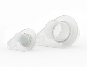 Reversible Strainers
Reversible strainer for filtration of single cells and isolation of cellular aggregates, including embryoid bodies and spheroids
-
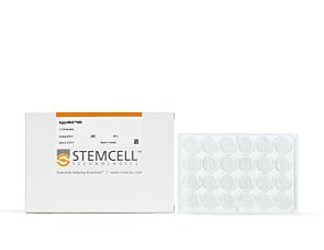 AggreWell™400
Microwell culture plates for easy and reproducible production of embryoid bodies and spheroids
-
 Y-27632 (Dihydrochloride)
RHO/ROCK pathway inhibitor; Inhibits ROCK1 and ROCK2
Overview
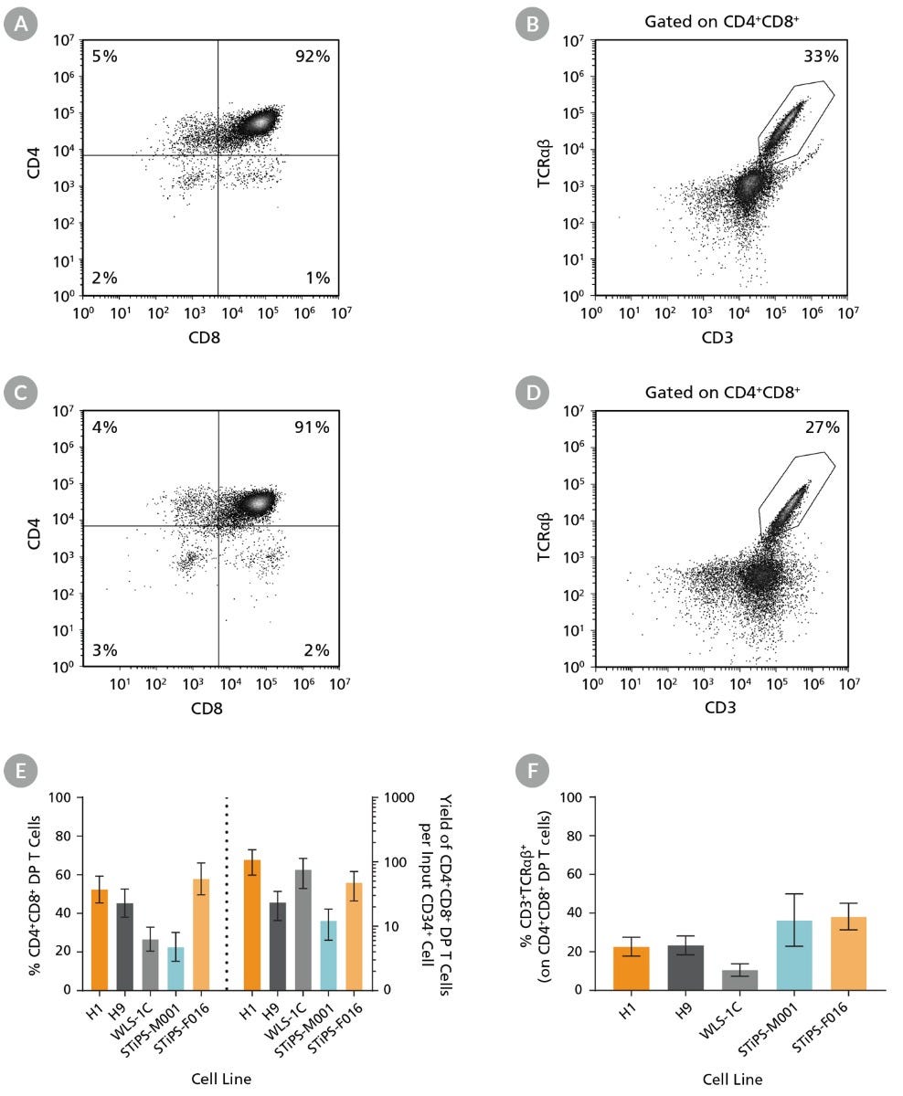
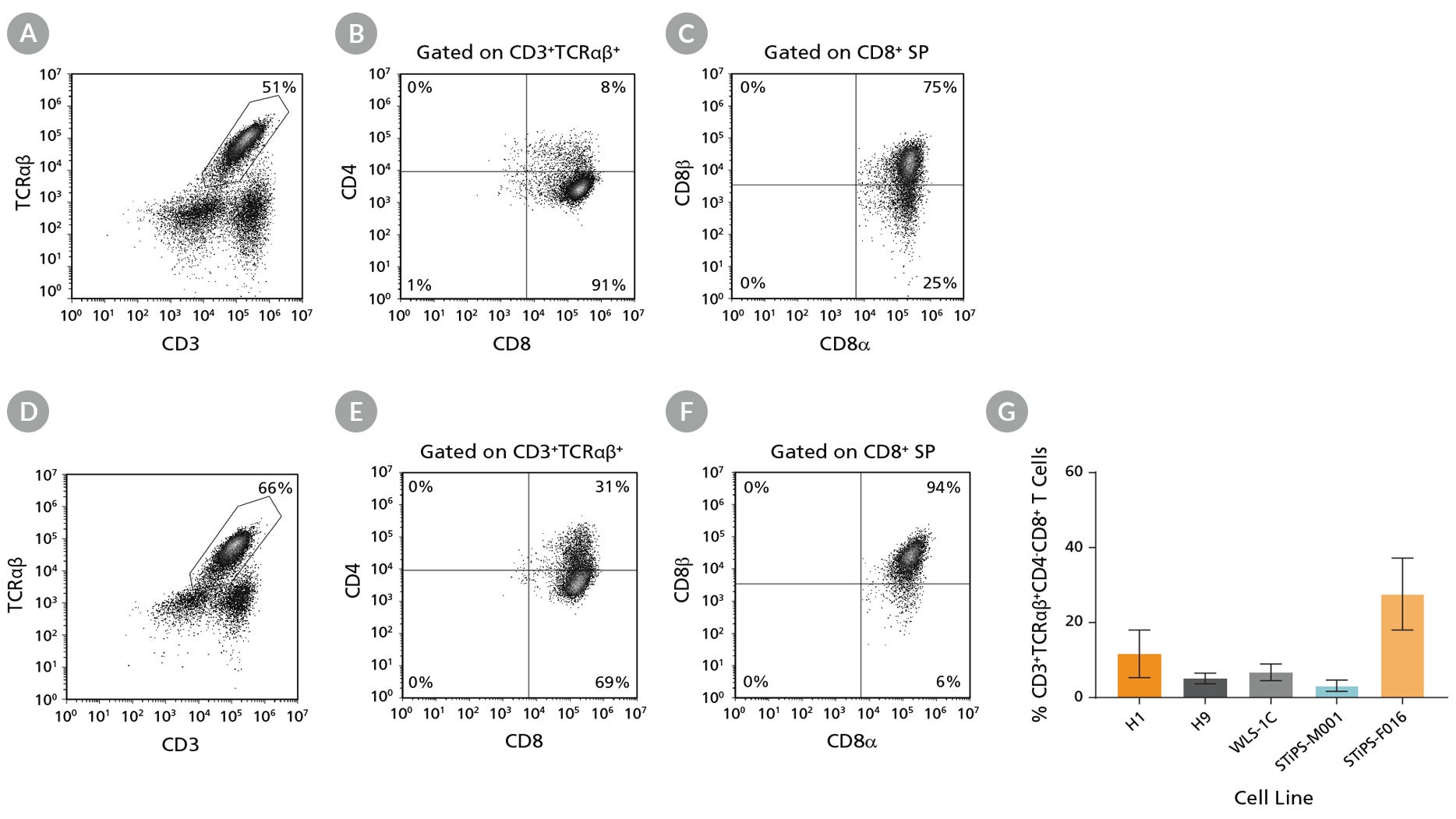
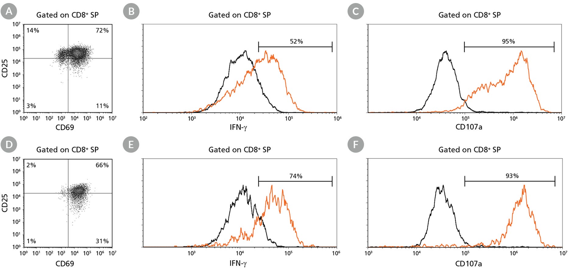
Data Figures
Protocols and Documentation
Find supporting information and directions for use in the Product Information Sheet or explore additional protocols below.
Applications
This product is designed for use in the following research area(s) as part of the highlighted workflow stage(s). Explore these workflows to learn more about the other products we offer to support each research area.
Resources and Publications
Educational Materials (12)


Related Products

PRODUCTS ARE FOR RESEARCH USE ONLY AND NOT INTENDED FOR HUMAN OR ANIMAL DIAGNOSTIC OR THERAPEUTIC USES UNLESS OTHERWISE STATED. FOR ADDITIONAL INFORMATION ON QUALITY AT STEMCELL, REFER TO WWW.STEMCELL.COM/COMPLIANCE.
