STEMdiff™ Mesenchymal Progenitor Kit

Defined culture kit for derivation and expansion of mesenchymal progenitor cells
STEMdiff™ Mesenchymal Progenitor Kit

Defined culture kit for derivation and expansion of mesenchymal progenitor cells

From: 841 USD
Catalog #
(Select a product)
Defined culture kit for derivation and expansion of mesenchymal progenitor cells
Add to Wish List

Product Advantages


  • Serum- and animal component-free formulation

  • Efficient and reproducible generation of MPCs from human ES and iPS cell lines

  • Rapid derivation of MPCs in 3 weeks

  • Generates MPCs capable of long-term expansion and differentiation to adipocytes, osteoblasts, and chondrocytes

What's Included

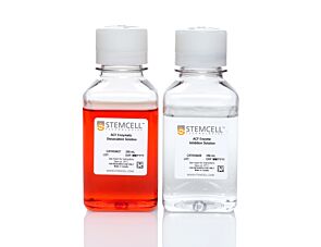
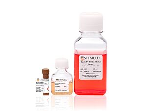
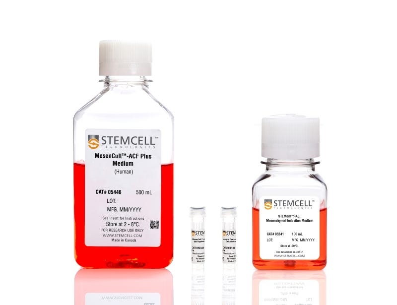
  • STEMdiff™-ACF Mesenchymal Induction Medium, 100 mL
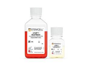
  • MesenCult™-ACF Plus Medium, 500 mL
  • MesenCult™-ACF Plus 500X Supplement, 1 mL
  • Animal Component-Free Cell Attachment Substrate, 1 mL
Products for Your Protocol
To see all required products for your protocol, please consult the Protocols and Documentation.

Overview

STEMdiff™ Mesenchymal Progenitor Kit is a defined culture kit consisting of animal component-free (ACF) induction medium, expansion medium, and attachment substrate. It is optimized for the derivation of cells with mesenchymal progenitor cell (MPC)-like properties from human embryonic stem (ES) cells or induced pluripotent stem (iPS) cells. This kit provides a complete workflow of defined reagents for derivation and expansion of human ES- or iPS-derived MPCs.

CollPlant is the manufacturer of the rhCollagen component of the cell attachment substrate.

The Product may be used for research purposes only. Please contact STEMCELL for any clinical or commercial application of the Product.
Subtype
Specialized Media
Cell Type
Mesenchymal Cells, PSC-Derived
Species
Human
Application
Cell Culture, Differentiation
Brand
STEMdiff
Area of Interest
Stem Cell Biology
Formulation Category
Animal Component-Free, Serum-Free

Data Figures

Human ES- and iPS-derived MPCs Can Be Further Differentiated Into Adipogenic, Chondrogenic and Osteogenic Lineages

Figure 1. Schematic of Differentiation Protocol and Timeline

In Phase 1, human ES or iPS cells are cultured in mTeSR™1 or TeSR™-E8™ medium. On Day 0 (Phase 2) of the protocol, cells are ready for induction into early mesoderm progenitor cells by replacing TeSR™ medium with STEMdiff™ Mesenchymal Induction Medium. By Day 4 (Phase 3), STEMdiff™ Mesenchymal Induction Medium is replaced with MesenCult™-ACF Medium to derive early mesenchymal progenitor cells (MPCs). On Day 6, cells are passaged onto cultureware precoated with MesenCult™-ACF Attachment Substrate in MesenCult™-ACF Medium. By Day 21, human ES- or iPS-derived MPCs exhibit the suggested MPC characteristics.

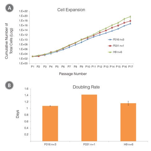
Cell Expansion and Doubling Rate of MPCs Derived from Human ES (H9) and iPS (STiPS-F016 and -F031) Cells in MesenCult™-ACF Medium

Figure 2. Cell Expansion and Doubling Rate of MPCs Derived from Human ES (H9) and iPS (STiPS-F016 and -F031) Cells in MesenCult™-ACF Medium

(A) The average cell expansion per passage over 17 passages for MPCs derived from human ES and iPS cell lines are approximately 9 and 10 fold. (B) Days to double cell number for human ES- and iPS-derived MPCs range from 1.1 to 1.4 days.

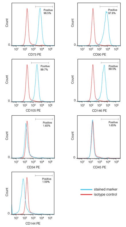
A Representative Flow Cytometric Analysis of STiPS-F016-derived MPCs Expressing Mesenchymal Surface Markers By Day 21

Figure 3. A Representative Flow Cytometric Analysis of STiPS-F016-derived MPCs Expressing Mesenchymal Surface Markers By Day 21

Human iPS-derived MPCs, generated using the STEMdiff™ Mesenchymal Progenitor Kit, express high levels of mesenchymal surface markers (CD73, CD90 and CD105) and the perivascular marker, CD146. MPCs do not express hematopoietic (CD34, CD45) and endothelial (CD144) surface markers. Human ES-derived MPCs express the same phenotype (data not shown).

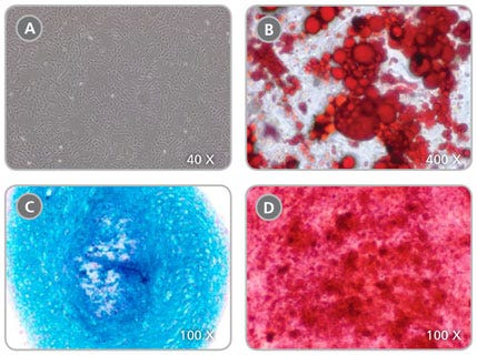
Human ES- and iPS-derived MPCs Can Be Further Differentiated Into Adipogenic, Chondrogenic and Osteogenic Lineages

Figure 4. Human ES- and iPS-derived MPCs Can Be Further Differentiated Into Adipogenic, Chondrogenic and Osteogenic Lineages

(A) MPCs generated from the 3 week protocol (described in Figure 1) and subsequently cultured in MesenCult™-ACF Medium develop MPC-like morphology (40X magnification). MPCs can be differentiated to (B) adipocytes (Oil Red O staining), 400X magnification; (C) chondrocytes (Alcian Blue staining), 100X magnification; and (D) osteoblasts (Fast Red and Silver Nitrate staining), 100X magnification.

Protocols and Documentation

Find supporting information and directions for use in the Product Information Sheet or explore additional protocols below.

Document Type
Product Name
Catalog #
Lot #
Language
Catalog #
05240
Lot #
All
Language
English
Document Type
Safety Data Sheet 1
Catalog #
05240
Lot #
All
Language
English
Document Type
Safety Data Sheet 2
Catalog #
05240
Lot #
All
Language
English
Document Type
Safety Data Sheet 3
Catalog #
05240
Lot #
All
Language
English
Document Type
Safety Data Sheet 4
Catalog #
05240
Lot #
All
Language
English

Applications

This product is designed for use in the following research area(s) as part of the highlighted workflow stage(s). Explore these workflows to learn more about the other products we offer to support each research area.

Resources and Publications

Publications (6)

Induced pluripotency and spontaneous reversal of cellular aging in supercentenarian donor cells. J. Lee et al. Biochemical and biophysical research communications 2020 may

Abstract

Supercentenarians (≥110-year-old, SC) are a uniquely informative population not only because they surpass centenarians in age, but because they appear to age more slowly with fewer incidences of chronic age-related disease than centenarians. We reprogramed donor B-lymphoblastoid cell lines (LCL) derived from a 114-year-old (SC), a 43-year-old healthy disease-free control (HDC) and an 8-year-old with a rapid aging disease (Hutchinson-Gilford progeria syndrome (HGPS)) and compared SC-iPSC to HDC-iPSC and HGPS-iPSCs. Reprogramming to pluripotency was confirmed by pluripotency marker expression and differentiation to 3 germ-layers. Each iPSC clone differentiated efficiently to mesenchymal progenitor cells (MPC) as determined by surface marker expression and RNAseq analysis. We identified supercentenarian and HGPS associated gene expression patterns in the differentiated MPC lines that were not evident in the parental iPSC lines. Importantly, telomere length resetting occurred in iPSC from all donors albeit at a lower incidence in supercentenarian iPSCs. These data indicate the potential to use reprogramming to reset both developmental state and cellular age in the oldest of the old." We anticipate that supercentenarian iPSC and their differentiated derivatives will be valuable tools for studying the underlying mechanisms of extreme longevity and disease resistance."
Intravenous administration of iPS-MSCSPIONs mobilized into CKD parenchyma and effectively preserved residual renal function in CKD rat. J.-J. Sheu et al. Journal of cellular and molecular medicine 2020 mar

Abstract

This study traced intravenously administered induced pluripotent stem cell (iPSC)-derived mesenchymal stem cells (MSC) and assessed the impact of iPSC-MSC on preserving renal function in SD rat after 5/6 nephrectomy. The results of in vitro study showed that FeraTrack™Direct contrast particles (ie intracellular magnetic labelling) in the iPSC-MSC (ie iPS-MSCSPIONs ) were clearly identified by Prussian blue stain. Adult-male SD rats (n = 40) were categorized into group 1 (SC), group 2 [SC + iPS-MSCSPIONs (1.0 × 10⁶ cells)/intravenous administration post-day-14 CKD procedure], group 3 (CKD), group 4 [CKD + iPS-MSCSPIONs (0.5 × 10⁶ cells)] and group 5 [CKD + iPS-MSCSPIONs (1.0 × 10⁶ cells)]. By day-15 after CKD induction, abdominal MRI demonstrated that iPS-MSCSPIONs were only in the CKD parenchyma of groups 4 and 5. By day 60, the creatinine level/ratio of urine protein to urine creatinine/kidney injury score (by haematoxylin and eosin stain)/fibrotic area (Masson's trichrome stain)/IF microscopic finding of kidney injury molecule-1 expression was lowest in groups 1 and 2, highest in group 3, and significantly higher in group 4 than in group 5, whereas IF microscopic findings of podocyte components (ZO-1/synaptopodin) and protein levels of anti-apoptosis ((Bad/Bcl-xL/Bcl-2) exhibited an opposite pattern to creatinine level among the five groups (all P {\textless} .0001). The protein expressions of cell-proliferation signals (PI3K/p-Akt/m-TOR, p-ERK1/2, FOXO1/GSK3$\beta$/p90RSK), apoptotic/DNA-damage (Bax/caspases8-10/cytosolic-mitochondria) and inflammatory (TNF-$\alpha$/TNFR1/TRAF2/NF-$\kappa$B) biomarkers displayed an identical pattern to creatinine level among the five groups (all P {\textless} .0001). The iPS-MSCSPIONs that were identified only in CKD parenchyma effectively protected the kidney against CKD injury.
Precision installation of a highly efficient suicide gene safety switch in human induced pluripotent stem cells. Z.-D. Shi et al. Stem cells translational medicine 2020 jul

Abstract

Human pluripotent stem cells including induced pluripotent stem cells (iPSCs) and embryonic stem cells hold great promise for cell-based therapies, but safety concerns that complicate consideration for routine clinical use remain. Installing a safety switch" based on the inducible caspase-9 (iCASP9) suicide gene system should offer added control over undesirable cell replication or activity. Previous studies utilized lentiviral vectors to integrate the iCASP9 system into T cells and iPSCs. This method results in random genomic insertion of the suicide switch and inefficient killing of the cells after the switch is "turned on" with a small molecule (eg AP1903). To improve the safety and efficiency of the iCASP9 system for use in iPSC-based therapy we precisely installed the system into a genomic safe harbor the AAVS1 locus in the PPP1R12C gene. We then evaluated the efficiencies of different promoters to drive iCASP9 expression in human iPSCs. We report that the commonly used EF1$\alpha$ promoter is silenced in iPSCs and that the endogenous promoter of the PPP1R12C gene is not strong enough to drive high levels of iCASP9 expression. However the CAG promoter induces strong and stable iCASP9 expression in iPSCs and activation of this system with AP1903 leads to rapid killing and complete elimination of iPSCs and their derivatives including MSCs and chondrocytes in vitro. Furthermore iPSC-derived teratomas shrank dramatically or were completely eliminated after administration of AP1903 in mice. Our data suggest significant improvements on existing iCASP9 suicide switch technologies and may serve as a guide to other groups seeking to improve the safety of stem cell-based therapies."