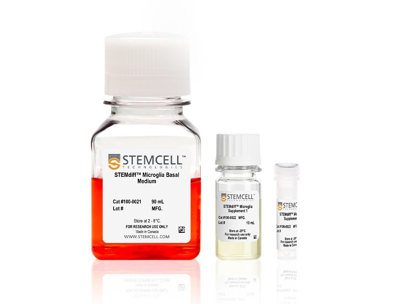
STEMdiff™ Microglia Differentiation Kit
Differentiation kit for the generation of microglia precursors from human ES and iPS cell-derived hematopoietic progenitor cells
Request Pricing
Thank you for your interest in this product. Please provide us with your contact information and your local representative will contact you with a customized quote. Where appropriate, they can also assist you with a(n):
Estimated delivery time for your area
Product sample or exclusive offer
In-lab demonstration
-
 STEMdiff™ Hematopoietic Kit
For differentiation of human ES or iPS cells into hematopoietic progenitor cells
-
 STEMdiff™ Microglia Maturation Kit
Maturation kit for the generation of microglia from human ES and iPS cell-derived microglia precursors
-
 DMEM/F-12 with 15 mM HEPES
Dulbecco's Modified Eagle's Medium/Nutrient Ham's Mixture F-12 (DMEM/F-12) with 15 mM HEPES buffer
-
 Falcon® Conical Tubes, 15 mL
Sterile polypropylene conical tubes for use in cell centrifugation and other cell culture applications
-
Labeling Antibodies
Compatible antibodies for purity assessment of isolated cells
Overview
Based on the protocol from the laboratory of Mathew Blurton-Jones (Abud et al., 2017), the resulting cells are a highly pure population of microglia (> 80% CD45/CD11b-positive, > 50% TREM2-positive microglia; < 20% morphologically distinct monocytes or macrophages).
Cells derived using these products are versatile tools for modeling neuroinflammation, studying human neurological development and disease, co-culture applications, and toxicity testing.
More Information
| Safety Statement | CA WARNING: This product can expose you to Progesterone which is known to the State of California to cause cancer. For more information go to www.P65Warnings.ca.gov |
|---|
Data Figures

Figure 1. Schematic for the STEMdiff™ Microglia Culture System Protocol
Microglial precursors can be generated in 24 days from hPSC-derived hematopoietic progenitor cells. For the generation of hematopoietic progenitor cells, see documentation for STEMdiff™ Hematopoietic Kit (Catalog #05310). For the maturation of microglial precursors to functional microglia, see the PIS.
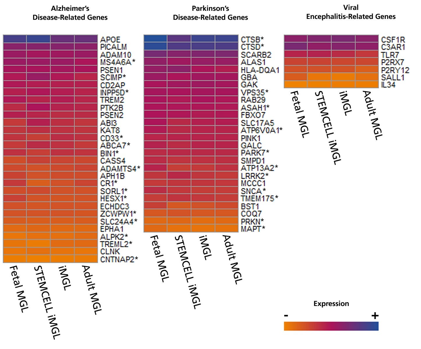
Figure 2. Microglia Generated with STEMdiff™ Microglia Culture System Express Disease-Relevant Genes Similar to Those from Published Differentiation and Maturation Protocols
Bulk RNA-seq datasets were extracted from 8 different publications that generated hPSC- (iMGL) and primary- (MGL) derived microglia and their transcriptional profiles compared to data from microglia generated with STEMdiff™ Microglia Culture System. The heat map displays absolute expression levels for select genes associated with Alzheimer’s disease, Parkinson’s disease, and viral encephalitis. Significant differences in gene expression between microglia generated with STEMdiff™ Microglia Culture System and any of the other 3 groups were identified by differential gene expression analysis. *= p<0.05 (DEseq2, adjusted). hPSC = human pluripotent stem cell.
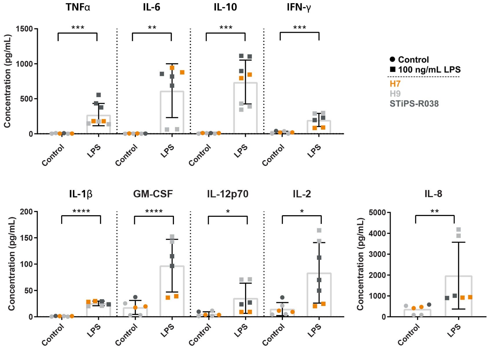
Figure 3. Microglia Generated with STEMdiff™ Microglia Culture System Release Cytokines in Response to Inflammatory Signals
Microglia were generated using the STEMdiff™ Microglia Culture System and stimulated with 100 ng/mL LPS for 24 hours. The release of pro-inflammatory (TNFα, IL-6, IFN-γ, IL-1β, GM-CSF, IL-12p70, IL-2, IL-8) and anti-inflammatory (IL-10) cytokines were measured by MSD. The microglia release cytokines in response to LPS treatment, as expected. *, p ≤ 0.05; **, p ≤ 0.01; ***, p ≤ 0.001; ****, p ≤ 0.0001. LPS = lipopolysaccharide; MSD = Meso Scale Discovery.
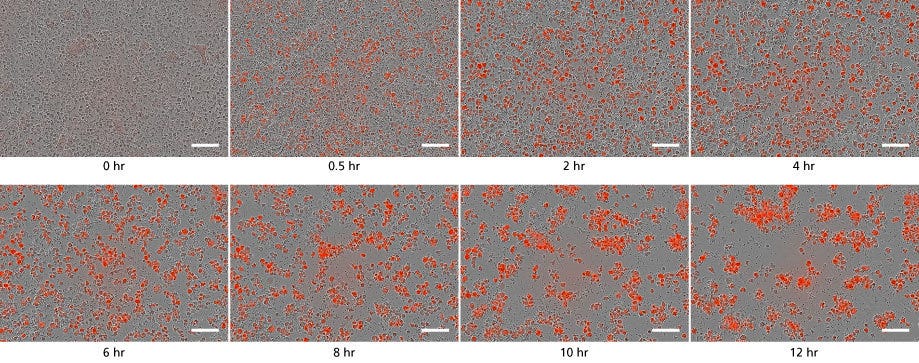
Figure 4. STEMdiff™ Microglia Culture System Generates Functional Microglia Capable of Phagocytosis at Day 34
Microglia taking up pH-sensitive bioindicator particles at a concentration of 250 μg/mL (small dots) were measured over a 12-hour time period with live cell imaging. As the particles are phagocytosed, the cells turn red. Over time, the number of small dots decreased, and the red cells increased in number and aggregated. Scale bar = 100μm.
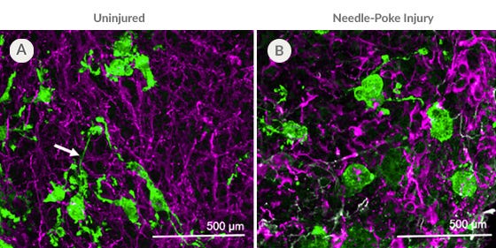
Figure 5. PSC-Derived Microglia Incorporate into Brain Organoids After 10 Days and Display an Activated Morphology upon Injury.
(A) Representative microglia and brain organoid co-cultures after 10 days, stained with IBA1 for microglia (green) and MAP2 for neurons (magenta). The microglia integrate among the neurons and display an unactivated morphology with extended processes (arrow). (B) The microglia display an activated amoeboid morphology upon injury as shown by IBA1 staining.
Protocols and Documentation
Find supporting information and directions for use in the Product Information Sheet or explore additional protocols below.
Applications
This product is designed for use in the following research area(s) as part of the highlighted workflow stage(s). Explore these workflows to learn more about the other products we offer to support each research area.
Resources and Publications
Educational Materials (27)
Related Products
-
 NeuroCult™ SM1 Neuronal Supplement
Supplement (50X) for the serum-free culture of neurons
-
 BrainPhys™ Neuronal Medium
Serum-free neurophysiological basal medium for improved neuronal function
-
 STEMdiff™ Cerebral Organoid Kit
Culture medium kit for establishment and maturation of human cerebral organoids
-
 STEMdiff™ Forebrain Neuron Differentiation ...
Differentiation kit for the generation of neuronal precursors from human ES and iPS cell-derived neural progenitor cells
Item added to your cart

STEMdiff™ Microglia Differentiation Kit
Copyright © 2024 by STEMCELL Technologies Inc. All rights reserved including graphics and images. STEMCELL Technologies & Design, STEMCELL Shield Design, Scientists Helping Scientists, and STEMdiff are trademarks of STEMCELL Technologies Canada Inc. All other trademarks are the property of their respective holders. While STEMCELL has made all reasonable efforts to ensure that the information provided by STEMCELL and its suppliers is correct, it makes no warranties or representations as to the accuracy or completeness of such information.
PRODUCTS ARE FOR RESEARCH USE ONLY AND NOT INTENDED FOR HUMAN OR ANIMAL DIAGNOSTIC OR THERAPEUTIC USES UNLESS OTHERWISE STATED. FOR ADDITIONAL INFORMATION ON QUALITY AT STEMCELL, REFER TO WWW.STEMCELL.COM/COMPLIANCE.
CA WARNING: This product can expose you to Progesterone which is known to the State of California to cause cancer. For more information go to www.P65Warnings.ca.gov


