STEMdiff™ Hematopoietic Kit

For differentiation of human ES or iPS cells into hematopoietic progenitor cells
Need a high-quality cell source? Choose from our hiPSC healthy control lines, manufactured with mTeSR™ Plus.

STEMdiff™ Hematopoietic Kit

For differentiation of human ES or iPS cells into hematopoietic progenitor cells

From: 829 USD
Catalog #
(Select a product)
For differentiation of human ES or iPS cells into hematopoietic progenitor cells
Add to Wish List

What's Included

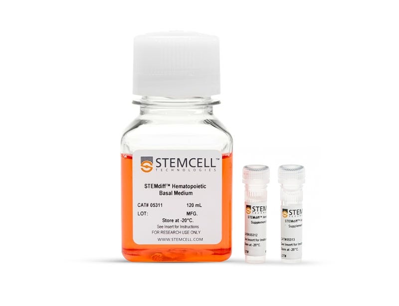
  • STEMdiff™ Hematopoietic Basal Medium, 120 mL
  • STEMdiff™ Hematopoietic Supplement A (200X), 225 µL
  • STEMdiff™ Hematopoietic Supplement B (200X), 375 µL

What Our Scientist Says

We developed the STEMdiff™ Hematopoietic Kit so scientists like you can have a reliable source of hPSC-derived hematopoietic progenitor cells for their research.

Marta WalasekSenior Scientist
Marta Walasek, Senior Scientist

Overview

Generate functional hematopoietic progenitor cells from human embryonic stem (ES) and induced pluripotent stem (iPS) cells with STEMdiff™ Hematopoietic Kit. Using a simple and reproducible protocol in serum- and feeder-free conditions, you can produce hematopoietic progenitor cells expressing CD34, CD45, and CD43.

The robust, two-stage protocol initially induces cells toward the mesoderm before further differentiating them into hematopoietic progenitor cells. After 12 days, a population of hematopoietic cells containing 25 - 65% (average 43%) CD34+CD45+ progenitor cells can be harvested, including progenitor cells that have the capacity to form functional hematopoietic colonies in the colony-forming unit (CFU) assay.

STEMdiff™ Hematopoietic Kit has been optimized for differentiation of human pluripotent cells maintained in:
• mTeSR™1 (Catalog #85850)
• mTeSR™ Plus (Catalog #100-0276)
• TeSR™-E8™ (Catalog #05990)
Subtype
Specialized Media
Cell Type
Hematopoietic Stem and Progenitor Cells, Pluripotent Stem Cells
Species
Human
Application
Cell Culture, Differentiation
Brand
STEMdiff
Area of Interest
Stem Cell Biology
Formulation Category
Serum-Free

Data Figures

Hematopoietic Cell Differentiation Protocol

Figure 1. Hematopoietic Differentiation Protocol

For 2D culture, start with harvesting and seeding human hESC/hiPSC colonies as small aggregates on Matrigel®-coated plates in mTeSR™1, mTeSR™ Plus, TeSR™-E8™ on Day -1, or in eTeSR™ on Day -2. For 3D culture, seed the aggregates in matrix-free suspension culture in TeSR™-AOF 3D on Day -1 using 6-well plates/Nalgene™ bottles on orbital shakers, or in PBS-MINI MagDrive Bioreactors. For both protocols, replace the TeSR™ medium with Medium A (STEMdiff™ Hematopoietic Basal Medium containing Supplement A) on Day 0 to begin inducing the cells toward a mesoderm-like state. On Day 2, a half-medium change is performed with fresh Medium A. On Day 3, the medium is changed to Medium B (STEMdiff™ Hematopoietic Basal Medium containing Supplement B) with half-medium changes on Days 5, 7 and 10, to promote further differentiation into hematopoietic cells. Typically, by Day 12, large numbers of HPCs can be harvested from the culture supernatant. For the hematopoietic differentiation protocol in 3D suspension culture, refer to this technical bulletin for step-by-step instructions.

Morphology of hPSC-Derived HPCs

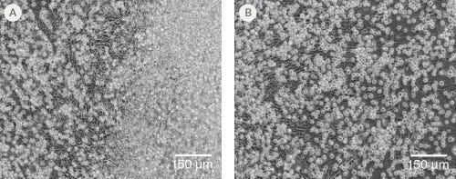
Figure 2. Morphology of hPSC-Derived HPCs

Representative images of (A) hES (H1) cells and (B) hiPS (WLS-1C) cells on Day 12 of differentiation to HPCs using the STEMdiff™ Hematopoietic Kit. Differentiated cells exhibit typical HPC morphology as round cells that float freely in suspension.

Efficient and Robust Generation of CD34+CD45+/CD43+ HPCs

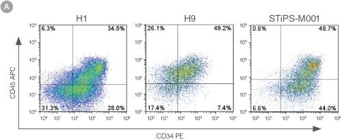
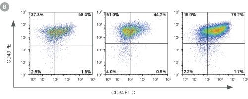
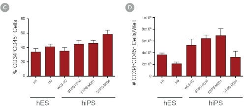
Figure 3. Efficient and Robust Generation of CD34+CD45+/CD43+ HPCs

hES and hiPS cells were cultured for 12 days in single wells of 12-well plates using the STEMdiff™ Hematopoietic Kit. At the end of the culture period, cells in suspension were harvested and analyzed by flow cytometry for expression of hematopoietic cell surface markers: CD34, CD45 and CD43. (A,B) Example flow cytometry plots for hematopoietic cell surface-marker analysis of cultures of hES (H1 and H9) and hiPS (STiPS-M001) cells. (C,D) Percentages and total numbers of CD34+CD45+ cells in cultures of hES (H1 and H9) or hiPS (WLS-1C, STiPS-F016, STiPS-M001 and STiPS-B004) cells are shown. Data shown as mean ± SEM; n ≥ 3.

hPSC-Derived HPCs Produce Colonies of Multiple Lineages

Figure 4. hPSC-Derived HPCs Produce Colonies of Multiple Lineages

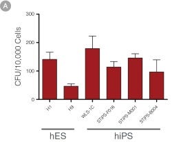
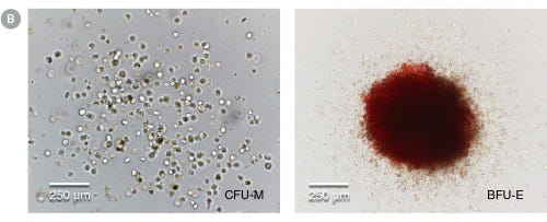
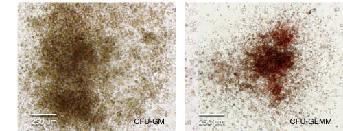
Cells in suspension were harvested from the cultures on Day 12 of the hematopoietic differentiation protocol and assessed in colony-forming unit (CFU) assays using MethoCult™ H4435 Enriched (Catalog #04435) methylcellulose-based medium. (A) CFU frequencies in cultures of 6 different hPSC lines. (Data shown as mean ± SEM; n ≥ 3.) The CFU frequencies were variable between the different cell lines, with on average approximately 120 CFU per 10,000 hPSC-derived HPCs plated. (B) The progenitor cell types observed included granulocyte/macrophage (CFU-M, CFU-G and CFU-GM), erythroid (BFU-E and CFU-E) and occasional mixed (CFU-GEMM) colonies. Representative colony images are shown at 40X magnification.

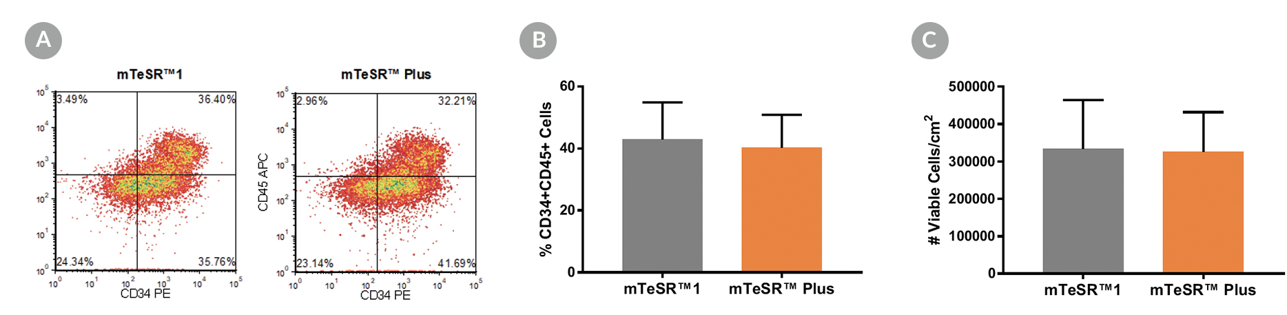
Density plots showing CD34+ and CD45+ expression and percentage of cells co-expressing CD34+ and CD45+ and graphs showing total number of viable cells harvested.

Figure 5. Generation of Hematopoietic Progenitor Cells from hPSCs Maintained in mTeSR™ Plus

Human ES (H1, H9) and iPS (STiPS-M001, WLS-1C) cell lines maintained in mTeSR™1 (daily feeds) or mTeSR™ Plus (restricted feeds) were differentiated to hematopoietic progenitor cells using the STEMdiff™ Hematopoietic Kit. At the end of the differentiation period, cells were harvested from the supernatant and analyzed by flow cytometry for co-expression of CD34+ and CD45+ . (A) Representative density plots showing CD34+ and CD45+ expression, (B) percentage of cells co-expressing CD34+ and CD45+ , and (C) total number of viable cells harvested are shown. Data are expressed as the mean (± SEM); n=4.

Protocols and Documentation

Find supporting information and directions for use in the Product Information Sheet or explore additional protocols below.

Document Type
Product Name
Catalog #
Lot #
Language
Catalog #
05310
Lot #
All
Language
English
Document Type
Safety Data Sheet 1
Catalog #
05310
Lot #
All
Language
English
Document Type
Safety Data Sheet 2
Catalog #
05310
Lot #
All
Language
English
Document Type
Safety Data Sheet 3
Catalog #
05310
Lot #
All
Language
English

Applications

This product is designed for use in the following research area(s) as part of the highlighted workflow stage(s). Explore these workflows to learn more about the other products we offer to support each research area.

Resources and Publications

Educational Materials (24)

Publications (40)

Innate Immunocompetent hiPSC-Derived Neurospheroids Capture Early CNS Responses to rAAV. C. Gomes et al. Advanced science (Weinheim, Baden-Wurttemberg, Germany) 2026 Sep

Abstract

Gene therapies using adeno-associated viruses (AAVs) for central nervous system (CNS) disorders face challenges because host immune responses are not represented in classical preclinical models. Here, we present a human-induced pluripotent stem cell (hiPSC)-derived innate immunocompetent 3D CNS model that recapitulates neuroinflammatory hallmarks, serving as a platform for preclinical gene therapy development. By utilizing various scales of stirred-tank bioreactor systems, we generated neurospheroids (iNSpheroids) composed of neurons, astrocytes, and oligodendrocytes, alongside microglial cells (iMGLs) to mimic the neuroimmune axis. These systems enabled large-scale production of iNSpheroids and subsequent miniaturization for co-culture experiments and screening of inflammatory stimuli, while maintaining a highly controlled environment. The iMGL-iNSpheroids demonstrated active neuron-microglia crosstalk and exhibited distinct inflammatory responses to a series of neuroinflammatory factors. iMGL-iNSpheroids mounted an early response to rAAV9, which is underscored by the activation of inflammatory pathways (e.g., TNF-via NF-κB activation) in glial cell populations. This model offers a valuable tool to dissect neuroinflammatory mechanisms, accelerating gene therapy development.
Cross-tissue immune profiling of APOE ε4 reveals early dysregulation in Alzheimer's disease. A. Shvetcov et al. Alzheimer's & dementia : the journal of the Alzheimer's Association 2026 Sep

Abstract

INTRODUCTION: Apolipoprotein E (APOE) ε4 is the strongest genetic risk factor for late-onset Alzheimer's disease (AD), but its contribution to disease pathogenesis remains incompletely understood. METHODS: Here, we integrate proteomic profiling of plasma (n = 9028), cerebrospinal fluid (n = 1099), dorsolateral prefrontal cortex (n = 720), and superior temporal gyrus (n = 105) to define the immune phenotype associated with APOE ε4. RESULTS: We identify a conserved, allele dose-dependent pro-inflammatory immune protein signature across peripheral and central tissues independent of AD diagnosis. This signature also emerges in patient-derived cortical organoids prior to amyloid beta and tau pathology, supporting a genotype-driven mechanism. Cross-tissue comparisons reveal shared innate and antiviral responses alongside tissue-specific immune signaling. Notably, a 12-week medical ketogenic diet partially reversed the APOE ε4 immune signature. DISCUSSION: These findings position immune dysregulation as an early and tractable driver of AD risk in APOE ε4 carriers with direct implications for targeted prevention strategies.
A human pluripotent stem cell triculture platform to elucidate microglial regulation of retinal ganglion cells in neuroinflammation. J. Harkin et al. Proceedings of the National Academy of Sciences of the United States of America 2026 Aug

Abstract

Optic neuropathies, including glaucoma, are characterized by the progressive degeneration of retinal ganglion cells (RGCs), ultimately leading to irreversible vision loss. Increasing evidence implicates microglia, the resident immune cells of the central nervous system, as key modulators of RGC health and disease progression. However, the precise mechanisms by which microglia influence RGCs remain poorly understood, particularly in the human context. In this study, we established human pluripotent stem cell-derived coculture systems incorporating microglia, astrocytes, and RGCs to explore how microglia shape RGC growth and maturation under physiological conditions. We first examined the impact of homeostatic microglia on RGCs in both coculture and triculture systems, revealing distinct influences of cell types in coculture compared to when they were grown individually. We then modeled inflammatory states by activating microglia with lipopolysaccharide and evaluated their effects on RGCs both directly and in the context of astrocyte coculture. This stepwise, reductionist approach enabled us to dissect the cellular interactions driving RGC vulnerability in inflammatory conditions relevant to optic neuropathies. Our findings provide insight into the complex neuroimmune landscape that underlies RGC degeneration and identify key pathways that may serve as therapeutic targets across a range of optic nerve diseases.
Need a high-quality cell source? Choose from our hiPSC healthy control lines, manufactured with mTeSR™ Plus.