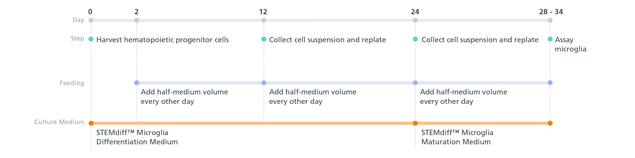
STEMdiff™ Microglia Differentiation Kit
Differentiation kit for the generation of microglia precursors from human ES and iPS cell-derived hematopoietic progenitor cells
Request Pricing
Thank you for your interest in this product. Please provide us with your contact information and your local representative will contact you with a customized quote. Where appropriate, they can also assist you with a(n):
Estimated delivery time for your area
Product sample or exclusive offer
In-lab demonstration
-
 STEMdiff™ Hematopoietic Kit
For differentiation of human ES or iPS cells into hematopoietic progenitor cells
-
 STEMdiff™ Microglia Maturation Kit
Maturation kit for the generation of microglia from human ES and iPS cell-derived microglia precursors
-
 DMEM/F-12 with 15 mM HEPES
Dulbecco's Modified Eagle's Medium/Nutrient Ham's Mixture F-12 (DMEM/F-12) with 15 mM HEPES buffer
-
 Falcon® Conical Tubes, 15 mL
Sterile polypropylene conical tubes for use in cell centrifugation and other cell culture applications
Overview
More Information
| Safety Statement | CA WARNING: This product can expose you to Progesterone which is known to the State of California to cause cancer. For more information go to www.P65Warnings.ca.gov |
|---|
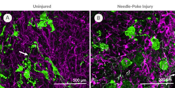
Data Figures
Protocols and Documentation
Find supporting information and directions for use in the Product Information Sheet or explore additional protocols below.
Applications
This product is designed for use in the following research area(s) as part of the highlighted workflow stage(s). Explore these workflows to learn more about the other products we offer to support each research area.
Resources and Publications
Educational Materials (27)


Related Products

Copyright © 2024 by STEMCELL Technologies Inc. All rights reserved including graphics and images. STEMCELL Technologies & Design, STEMCELL Shield Design, Scientists Helping Scientists, and STEMdiff are trademarks of STEMCELL Technologies Canada Inc. All other trademarks are the property of their respective holders. While STEMCELL has made all reasonable efforts to ensure that the information provided by STEMCELL and its suppliers is correct, it makes no warranties or representations as to the accuracy or completeness of such information.
PRODUCTS ARE FOR RESEARCH USE ONLY AND NOT INTENDED FOR HUMAN OR ANIMAL DIAGNOSTIC OR THERAPEUTIC USES UNLESS OTHERWISE STATED. FOR ADDITIONAL INFORMATION ON QUALITY AT STEMCELL, REFER TO WWW.STEMCELL.COM/COMPLIANCE.
CA WARNING: This product can expose you to Progesterone which is known to the State of California to cause cancer. For more information go to www.P65Warnings.ca.gov
