BrainPhys™ Neuronal Medium N2-A & SM1 Kit

Kit for serum-free culture of ES/iPS cell-derived neurons in BrainPhys™ Neuronal Medium
BrainPhys™ Neuronal Medium N2-A & SM1 Kit

Kit for serum-free culture of ES/iPS cell-derived neurons in BrainPhys™ Neuronal Medium

From: 388 USD
Catalog #
(Select a product)
Kit for serum-free culture of ES/iPS cell-derived neurons in BrainPhys™ Neuronal Medium
Add to Wish List

Product Advantages


  • More representative of the brain’s extracellular environment

  • Improved neuronal function and a higher proportion of synaptically active neurons

  • Perform functional assays without changing media and shocking cells

  • Supports long-term culture of ES/iPS cell- and CNS-derived neurons

  • Rigorous raw material screening and quality control ensure minimal lot-to-lot variability

What's Included

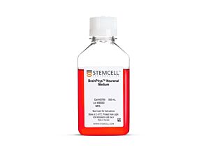
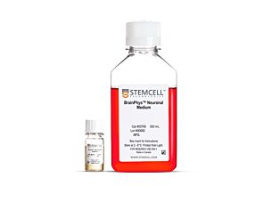
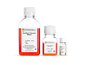
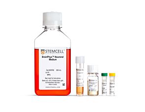
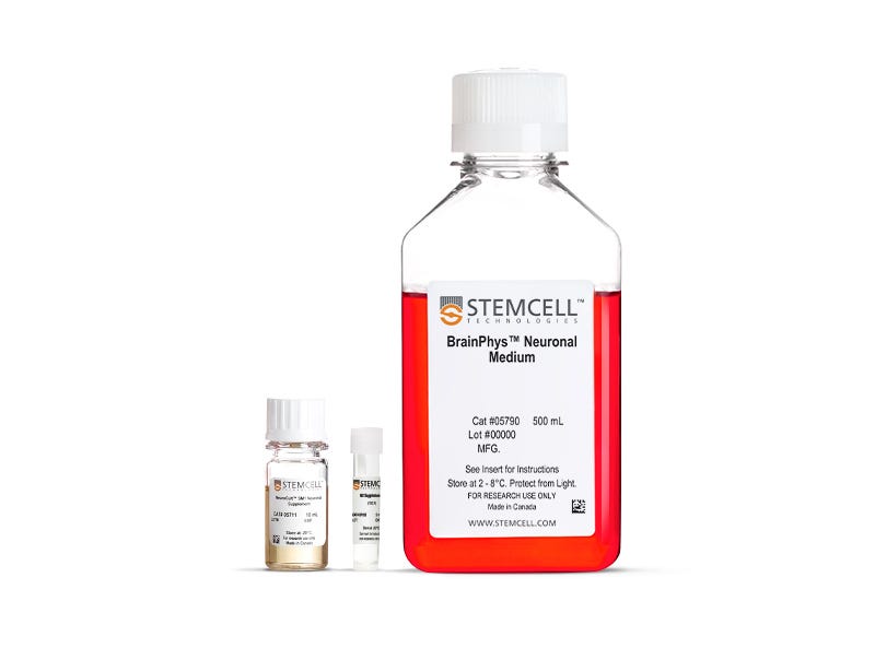
  • BrainPhys™ Neuronal Medium, 500 mL (Catalog #05790)
  • NeuroCult™ SM1 Neuronal Supplement, 10 mL (Catalog #05711)
  • N2 Supplement-A, 5 mL (Catalog #07152)

What Our Scientist Says

I want to help neuroscientists like you create more physiological culture conditions, for more active and healthy neuronal cultures.

Carmen MakScientist
Carmen Mak, Scientist

Overview

Culture, differentiate, and mature neurons derived from human embryonic stem (ES) or induced pluripotent stem (iPS) cells in a complete medium optimized to promote, rather than inhibit neuronal activity.

For your convenience, BrainPhys™ Neuronal Medium N2-A & SM1 Kit includes BrainPhys™ Neuronal Medium (basal medium) and supplements for culturing human ES/iPS-derived neuronal progenitor cells. Based on the formulation by Bardy and Gage (Bardy et al. PNAS, 2015), serum-free BrainPhys™ Neuronal Medium mimics the extracellular environment of the central nervous system (CNS) to yield a higher proportion of synaptically active neurons. Brewer’s B27-based (Brewer et al. J Neurosci Res., 1993) NeuroCult™ SM1 Neuronal Supplement ensures cell health and encourages neurite outgrowth and branching in short- and long-term serum-free cultures, and N2 Supplement-A supports the in vitro differentiation of ES/iPS-derived cells to neuronal subtypes. For lineage-specific differentiation requiring growth factors, please see BrainPhys™ hPSC Neuron Kit, which also includes BDNF and GDNF.

To avoid shocking your cells with media changes, you can also use BrainPhys™ medium when performing functional assays, such as microelectrode array-based recordings or live-fluorescent imaging.

View our additional resources to learn more about the BrainPhys™system.
Subtype
Basal Media, Specialized Media
Cell Type
Neural Cells, PSC-Derived, Neurons, Pluripotent Stem Cells
Species
Human
Application
Cell Culture, Differentiation, Maintenance
Brand
BrainPhys
Area of Interest
Disease Modeling, Drug Discovery and Toxicity Testing, Neuroscience, Stem Cell Biology
Formulation Category
Serum-Free

Data Figures

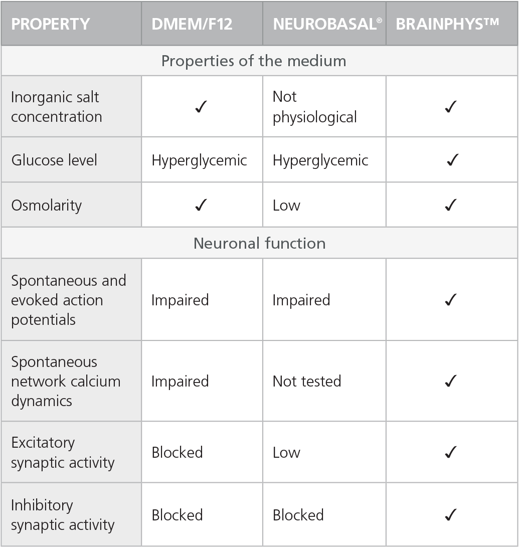
Table 1. Properties of Culture Media (C Bardy et al. Proc Natl Acad Sci USA, 2015)

Check-mark denotes physiological conditions

Check-mark denotes physiological conditions and supported activities according to C Bardy et al. Proc Natl Acad Sci USA, 2015.

Rodent Neurons Matured in BrainPhys™ Neuronal Medium

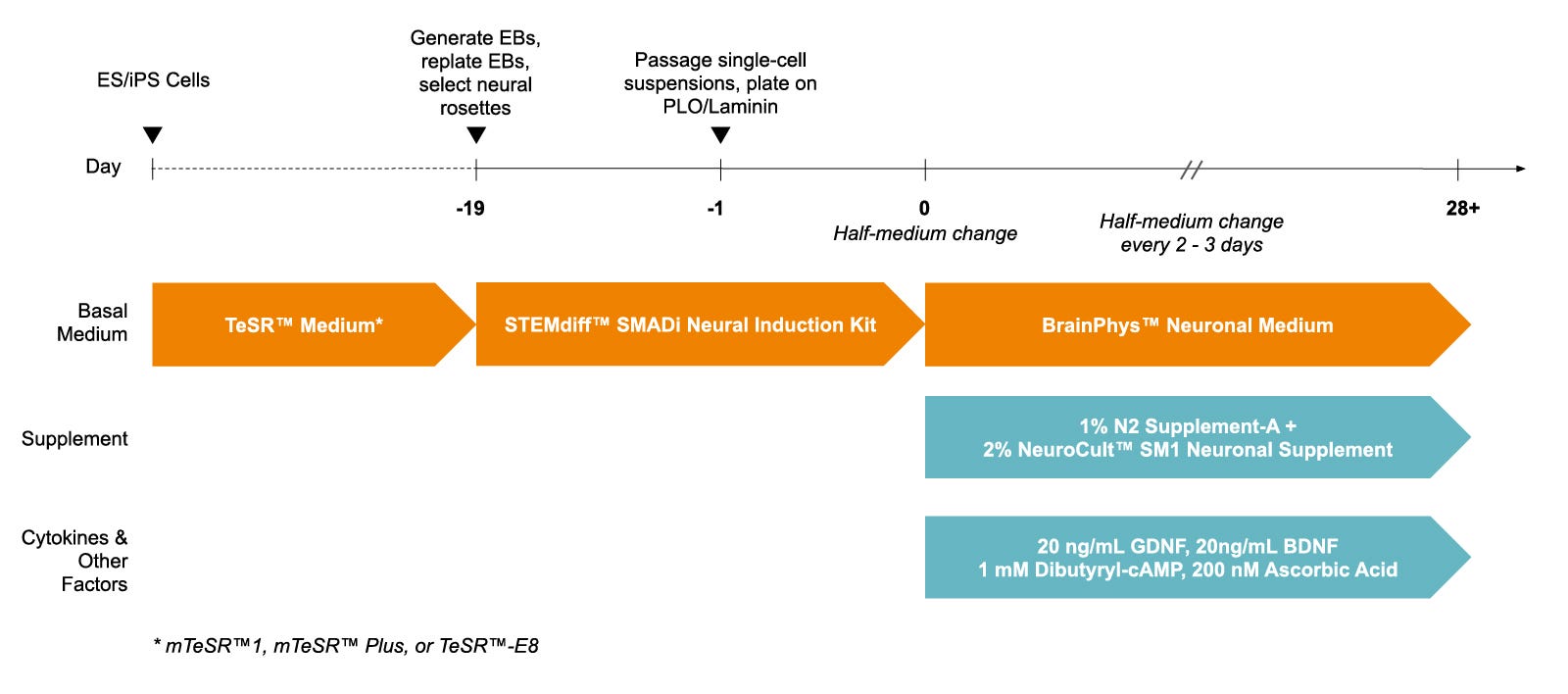
Figure 1. Protocol for Culturing hPSCs with the SM1 Culture System

hPSCs were maintained in mTeSR™1 medium and then differentiated using the STEMdiff™ SMADi Neural Induction Kit. Following plating on PLO/laminin, half-medium changes were performed to transition to BrainPhys™ Neuronal Medium for maturation and long-term culture.

hPSC-Derived Neurons Generated in BrainPhys™ Neuronal Medium and NeuroCult™ SM1 and N2 Supplements are Healthy and Morphologically Normal

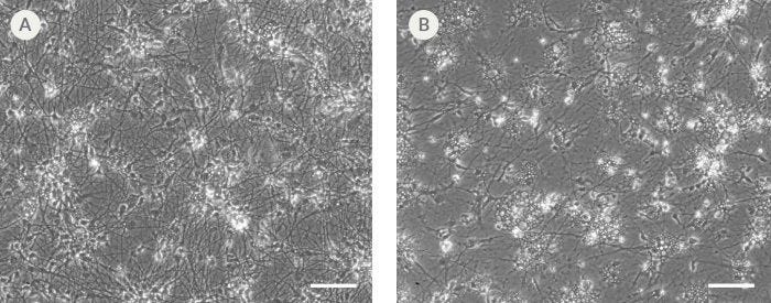
Figure 2. hPSC-Derived Neurons Generated in BrainPhys™ Neuronal Medium and NeuroCult™ SM1 and N2 Supplements are Healthy and Morphologically Normal

NPCs were generated from H9 cells using STEMdiff™ Neural Induction Medium in an embryoid body-based protocol. Next, NPCs were cultured for 44 DIV in (A) BrainPhys™ Neuronal Medium, supplemented with 2% NeuroCult™ SM1 Supplement, 1% N2 Supplement-A, 20 ng/mL GDNF, 20 ng/mL BDNF, 1 mM db-cAMP and 200 nM ascorbic acid to initiate neuronal differentiation, or (B) DMEM/F12 under the same supplementation conditions. Neuronal cultures differentiated from NPCs in BrainPhys™ Neuronal Medium display extensive neurite outgrowth and reduced cellular debris compared to cultures differentiated in DMEM/F12. Scale bar= 100 µm.

Protocols and Documentation

Find supporting information and directions for use in the Product Information Sheet or explore additional protocols below.

Document Type
Product Name
Catalog #
Lot #
Language
Catalog #
05793
Lot #
All
Language
English
Document Type
Safety Data Sheet 1
Catalog #
05793
Lot #
All
Language
English
Document Type
Safety Data Sheet 2
Catalog #
05793
Lot #
All
Language
English
Document Type
Safety Data Sheet 3
Catalog #
05793
Lot #
All
Language
English

Applications

This product is designed for use in the following research area(s) as part of the highlighted workflow stage(s). Explore these workflows to learn more about the other products we offer to support each research area.

Resources and Publications

Educational Materials (22)

Publications (54)

NeuroSuite for Long-Term Functional and Structural Studies of Air-Liquid Interface Cerebral Organoids. B. Haider et al. Advanced science (Weinheim, Baden-Wurttemberg, Germany) 2026 Aug

Abstract

Long-term electrophysiological monitoring of human cerebral organoids remains challenging, as 3D tissues rapidly develop necrotic cores once diffusion limits are exceeded. Air-liquid interface cerebral organoids (ALI-COs) overcome this by maintaining thin slices with continuous metabolic exchange, preserving viability, yet no system allows stable, in situ recordings over months. Here, we introduce NeuroSuite, a modular platform for long-term, spatially resolved neural monitoring of ALI-COs. Its core, Neuroweb, is an ultrathin, perforated microelectrode array that preserves cellular diversity, evidenced by immunofluorescence and RNA sequencing, while enabling low-impedance (∼25 kΩ at 1 kHz) recordings. Complementing this, NeuroMaps, our open-source analysis suite, streamlines signal quality control, spike and field potential analysis, and integration with imaging. NeuroSuite is suitable for capturing maturation-dependent network dynamics, including potential excitation-inhibition shifts and oscillatory activity, beyond 180 days. Acute ex vivo rat brain slice recordings further demonstrate its versatility, establishing NeuroSuite as a robust platform for developmental neuroscience, disease modelling, and drug screening.
Generation of 3D Human iPSC-Derived Multi-Cell Type Neurospheres for Studying Neuron, Astrocyte, and Microglia Crosstalk S. Wendt et al. Bio-protocol 2025 Nov

Abstract

Three-dimensional (3D) human brain tissue models derived from induced pluripotent stem cells (iPSCs) have transformed the study of neural development and disease in vitro. While cerebral organoids offer high structural complexity, their large size often leads to necrotic core formation, limiting reproducibility and challenging the integration of microglia. Here, we present a detailed, reproducible protocol for generating multi-cell type 3D neurospheres that incorporate neurons, astrocytes, and optionally microglia, all derived from the same iPSCs. While neurons and astrocytes differentiate spontaneously from neural precursor cells, generated by dual SMAD-inhibition (blocking BMP and TGF-b signaling), microglia are generated in parallel and can infiltrate the mature neurosphere tissue after plating neurospheres into 48-well plates. The system supports a range of downstream applications, including functional confocal live imaging of GCaMP6f after adeno-associated virus (AAV) transduction of neurospheres or immunofluorescence staining after fixation. Our approach has been successfully implemented across multiple laboratories, demonstrating its robustness and translational potential for studying neuron–glia interactions and modeling neurodegenerative processes.
Loss?of?function variants in RNA binding motif protein X?linked induce neuronal defects contributing to amyotrophic lateral sclerosis pathogenesis MedComm 2024 Sep

Abstract

AbstractDespite being one of the most prevalent RNA modifications, the role of N6?methyladenosine (m6A) in amyotrophic lateral sclerosis (ALS) remains ambiguous. In this investigation, we explore the contribution of genetic defects of m6A?related genes to ALS pathogenesis. We scrutinized the mutation landscape of m6A genes through a comprehensive analysis of whole?exome sequencing cohorts, encompassing 508 ALS patients and 1660 population?matched controls. Our findings reveal a noteworthy enrichment of RNA binding motif protein X?linked (RBMX) variants among ALS patients, with a significant correlation between pathogenic m6A variants and adverse clinical outcomes. Furthermore, Rbmx knockdown in NSC?34 cells overexpressing mutant TDP43Q331K results in cell death mediated by an augmented p53 response. Similarly, RBMX knockdown in ALS motor neurons derived from induced pluripotent stem cells (iPSCs) manifests morphological defects and activation of the p53 pathway. Transcriptional analysis using publicly available single?cell sequencing data from the primary motor cortex indicates that RBMX?regulated genes selectively influence excitatory neurons and exhibit enrichment in ALS?implicated pathways. Through integrated analyses, our study underscores the emerging roles played by RBMX in ALS, suggesting a potential nexus between the disease and dysregulated m6A?mediated mRNA metabolism. The dysregulation of m6A modification has gained recognition as a crucial factor in the development of amyotrophic lateral sclerosis (ALS). Among the m6A reader proteins, RNA binding motif protein X?linked (RBMX) stands out with a notable enrichment of variants in ALS patients, and the presence of pathogenic RBMX variants is associated with a faster disease progression. In vitro experiments have provided evidence that reducing RBMX levels can result in neuronal defects. Additionally, bioinformatic analyses have supported these findings by revealing that RBMX?associated genes specifically impact excitatory neurons. Furthermore, these genes are involved in the regulation of pathways and genes associated with neurodegeneration and RNA metabolism, underscoring the relevance of RBMX in ALS pathogenesis.