Make more informed purchasing decisions with our new product availability and delivery estimate feature, now available on all product pages, in your cart, and during checkout.
Sign In
New to STEMCELL?
Register for an account to quickly and easily purchase products online and for one-click access to all educational content.
Thank you for your interest in this product.
Please provide us with your contact information and your local representative
will contact you with a customized quote. Where appropriate, they can also assist you with a(n):
Estimated delivery time for your area
Product sample or exclusive offer
In-lab demonstration
By submitting this form, you are providing your consent to STEMCELL Technologies Canada Inc. and its subsidiaries and affiliates (“STEMCELL”) to collect and use your information, and send you newsletters and emails in accordance with our privacy policy. Please contact us with any questions that you may have. You can unsubscribe or change your email preferences at any time.
The 2E1.E9 antibody reacts with glial fibrillary acidic protein (GFAP), an ~49 kDa type III intermediate filament (IF) protein that, within the central nervous system, is expressed primarily by astrocytes, though found at high levels in some glial-derived tumors. GFAP is thought to contribute to the structural architecture and strength of the cytoskeleton. The 2E1.E9 antibody does not cross-react with other IF proteins and can be used to distinguish astrocytes from other glial cells. GFAP has also been identified in Leydig cells, keratinocytes, chondrocytes and osteocytes. The GFAP polypeptide comprises an N-terminal head, a central rod, and a C-terminal tail domain, and assembles as dimers by a process dependent on phosphorylation and dephosphorylation of the N-terminal domain. Several splice variants have been identified, encoding three distinct isoforms. Many mutations in the GFAP gene (>50) have been associated with Alexander disease, a progressive leukoencephalopathy characterized by cytoplasmic inclusions and dysfunctional myelination.
This antibody clone has been verified for labeling neural stem and progenitor cells grown in NeuroCult™ NS-A Proliferation Kit (Human; Catalog #05751) and NeuroCult™ Proliferation Kit (Mouse; Catalog #05702).
Subtype
Primary Antibodies
Target Antigen
GFAP (Glial Fibrillary Acidic Protein)
Alternative Names
Glial fibrillary acid protein (GFAP)
Reactive Species
Human, Mouse, Rat
Conjugation
Unconjugated
Host Species
Mouse
Cell Type
Astrocytes, Neural Cells, PSC-Derived
Species
Human, Mouse, Rat
Application
Flow Cytometry, Immunocytochemistry, Immunofluorescence, Immunohistochemistry, Western Blotting
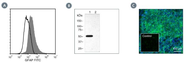
(A) Flow cytometry analysis of Sprague-Dawley rat brain cells labeled with Anti-GFAP Antibody, Clone 2E1.E9, followed by Goat Anti-Mouse IgG (H+L) Antibody, Polyclonal, FITC (Catalog #60138FI) (filled histogram), or a mouse IgG2b, kappa isotype control antibody, followed by Goat Anti-Mouse IgG (H+L) Antibody, Polyclonal, FITC (solid line histogram).
(B) Western blot analysis of denatured/reduced Sprague-Dawley rat brain lysate (lane 1) or HT1080 fibrosarcoma cells (negative control, lane 2) with Anti-GFAP Antibody, Clone 2E1.E9.
(C) Embryonic mouse cortical tissue was cultured using the NeuroCult™ Proliferation Kit (Mouse), then fixed and labeled with Anti-GFAP Antibody, Clone 2E1.E9, followed by goat anti-mouse IgG, FITC. Nuclei were counter-stained with DAPI. Inset shows cells labeled with a mouse IgG2b, kappa isotype control antibody, followed by goat anti-mouse IgG, FITC (without DAPI staining).
This product is designed for use in the following research area(s) as part
of the highlighted workflow stage(s). Explore these workflows to learn more about the other products we
offer to support each research area.
Onset of rosette formation during spontaneous neural differentiation of hESC and hiPSC colonies
Malchenko S et al.
Gene 2014 JAN
Abstract
In vitro neural differentiation of human embryonic stem cells (hESCs) is an advantageous system for studying early neural development. The process of early neural differentiation in hESCs begins by initiation of primitive neuroectoderm, which is manifested by rosette formation, with consecutive differentiation into neural progenitors and early glial-like cells. In this study, we examined the involvement of early neural markers - OTX2, PAX6, Sox1, Nestin, NR2F1, NR2F2, and IRX2 - in the onset of rosette formation, during spontaneous neural differentiation of hESC and human induced pluripotent stem cell (hiPSC) colonies. This is in contrast to the conventional way of studying rosette formation, which involves induction of neuronal differentiation and the utilization of embryoid bodies. Here we show that OTX2 is highly expressed at the onset of rosette formation, when rosettes comprise no more than 3-5 cells, and that its expression precedes that of established markers of early neuronal differentiation. Importantly, the rise of OTX2 expression in these cells coincides with the down-regulation of the pluripotency marker OCT4. Lastly, we show that cells derived from rosettes that emerge during spontaneous differentiation of hESCs or hiPSCs are capable of differentiating into dopaminergic neurons in vitro, and into mature-appearing pyramidal and serotonergic neurons weeks after being injected into the motor cortex of NOD-SCID mice. ?? 2013 Elsevier B.V.
Mouse monoclonal IgG2b, kappa isotype control antibody
Item added to your cart
Anti-GFAP Antibody, Clone 2E1.E9
Quality Statement:
PRODUCTS ARE FOR RESEARCH USE ONLY AND NOT INTENDED FOR HUMAN OR ANIMAL DIAGNOSTIC OR THERAPEUTIC USES UNLESS OTHERWISE STATED. FOR ADDITIONAL INFORMATION ON QUALITY AT STEMCELL, REFER TO WWW.STEMCELL.COM/COMPLIANCE.