STEMdiff™ Cerebral Organoid Kit

Culture medium kit for establishment and maturation of human cerebral organoids
Need a high-quality cell source? Choose from our hiPSC healthy control lines, manufactured with mTeSR™ Plus.

STEMdiff™ Cerebral Organoid Kit

Culture medium kit for establishment and maturation of human cerebral organoids

From: 470 USD
Catalog #
(Select a product)
Culture medium kit for establishment and maturation of human cerebral organoids
Add to Wish List

What's Included

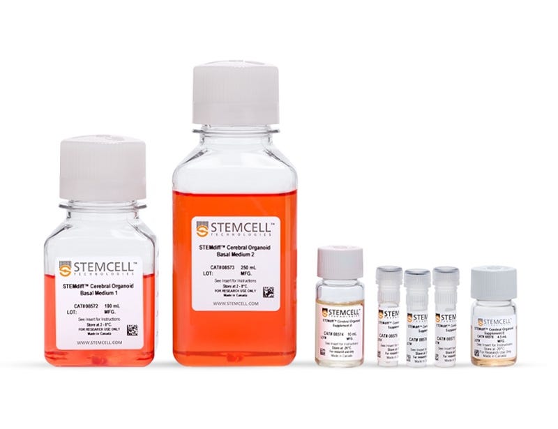
  • STEMdiff™ Cerebral Organoid Basal Medium 1,100 mL
  • STEMdiff™ Cerebral Organoid Basal Medium 2, 250 mL
  • STEMdiff™ Cerebral Organoid Supplement A, 10 mL
  • STEMdiff™ Cerebral Organoid Supplement B, 0.5 mL
  • STEMdiff™ Cerebral Organoid Supplement C, 0.25 mL
  • STEMdiff™ Cerebral Organoid Supplement D, 0.5 mL
  • STEMdiff™ Cerebral Organoid Supplement E, 4.5 mL
Products for Your Protocol
To see all required products for your protocol, please consult the Protocols and Documentation.

What Our Scientist Says

Human brain development is complex, so to observe it in a dish is a huge scientific breakthrough. We've tried to simplify that process and make it more accessible to you, regardless of how much stem cell experience you have.

Leon ChewScientist
Leon Chew, Scientist

Overview

Generate self-organized, pluripotent stem cell (PSC)-derived neural organoids with a cellular composition and structural organization representative of the developing human brain.

These defined, serum-free cell culture media and the simple, four-stage protocol are based on the formulation published by Lancaster et al. (Lancaster MA et al. Nature, 2013 and Lancaster MA et al. Science, 2014) to more reliably generate cerebral organoids. Beginning with an embryoid body (EB) formation step followed by expansion of neuroepithelia, organoids generated using STEMdiff™ Cerebral Organoid Kit feature cortical-like regions, including the ventricular zone (PAX6+/SOX2+/Ki-67+), outer subventricular zone (Ki-67+/p-Vimentin+), intermediate zone (TBR2+), and cortical plate (CTIP2+/MAP2+/TBR1+), which layer in similar orientations as those observed in vivo.

For extended culture periods (> 40 days), the components required for maturation can be purchased as STEMdiff™ Cerebral Organoid Maturation Kit.
Subtype
Specialized Media
Cell Type
Neural Cells, PSC-Derived, Neural Stem and Progenitor Cells, Pluripotent Stem Cells
Species
Human
Application
Cell Culture, Characterization, Differentiation, Functional Assay, Immunofluorescence, Organoid Culture, Phenotyping, Spheroid Culture
Brand
STEMdiff
Area of Interest
Disease Modeling, Drug Discovery and Toxicity Testing, Neuroscience, Stem Cell Biology
Formulation Category
Serum-Free

Data Figures

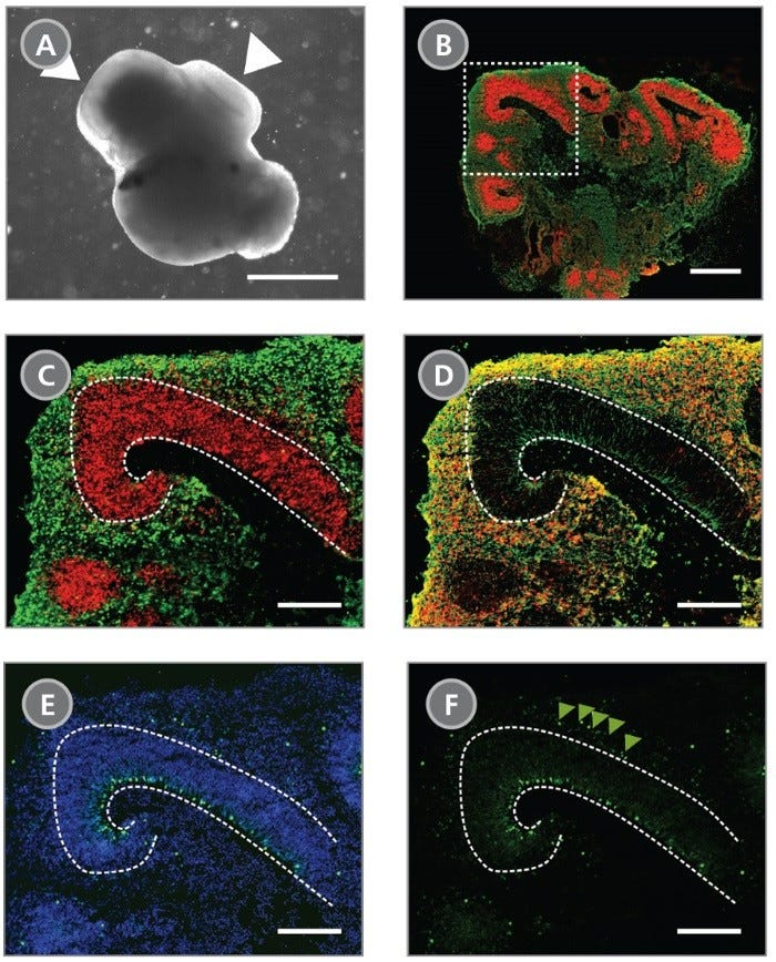
Cerebral Organoids Contain Multiple Layered Regions That Recapitulate the Cortical Lamination Process Observed During In Vivo Human Brain Development

Figure 1. Cerebral Organoids Contain Multiple Layered Regions That Recapitulate the Cortical Lamination Process Observed During In Vivo Human Brain Development

(A) A representative phase-contrast image of a whole cerebral organoid at Day 40 generated using the STEMdiff™ Cerebral Organoid Kit. Cerebral organoids at this stage are made up of phase-dark structures that may be surrounded by regions of thinner, more translucent structures that display layering (arrowheads). (B) Immunohistological analysis on cryosections of cerebral organoids reveals cortical regions within the organoid labeled by the apical progenitor marker PAX6 (red) and neuronal marker β-tubulin III+ (TUJ-1) (green). (C-F) Inset of boxed region from (B). (C) PAX6+ apical progenitors (red, enclosed by dotted line) are localized to a ventricular zone-like region. β-tubulin III+ neurons (green) are adjacent to the ventricular zone. (D) CTIP2, a marker of the developing cortical plate, co-localizes with β-tubulin III+ neurons in a cortical plate-like region. Organization of the layers recapitulates early corticogenesis observed during human brain development. (E) Proliferating progenitor cells labeled by Ki-67 (green) localize along the ventricle, nuclei are counterstained with DAPI (blue). (F) An additional population of Ki-67+ cells is found in an outer subventricular zone-like region (arrowheads). Scale bar = (A) 1 mm, (B) 1 mm and (C-F) 200 µm.

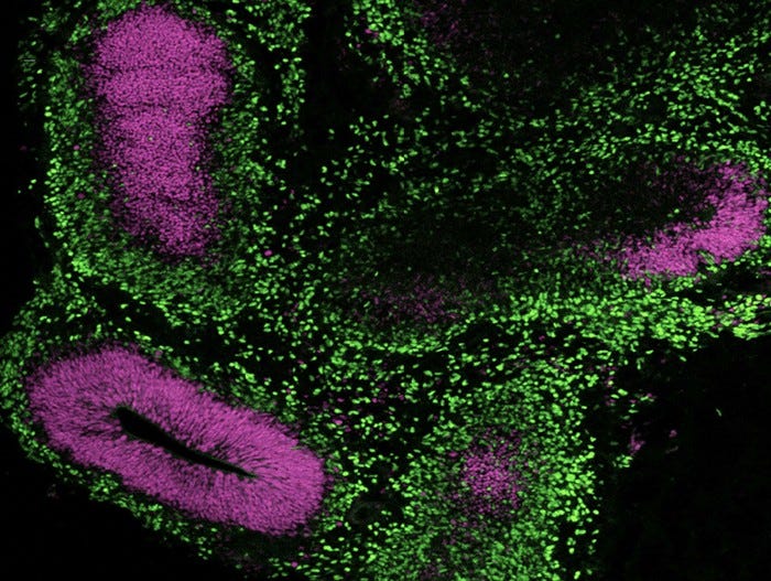
Immunocytochemistry image of a cerebral organoid cultured in mTeSR™ Plus and directed to cerebral organoids using the STEMdiff™ Cerebral Organoid Kit.

Figure 2. Cerebral Organoids Can Be Generated from hPSCs Maintained in mTeSR™ Plus

Human ES (H9) cells were cultured with mTeSR™ Plus and directed to cerebral organoids using the STEMdiff™ Cerebral Organoid Kit. Image shows apical progenitor marker SOX2 (magenta) and neuronal marker TBR1 (green).

Cryosectioned Cerebral Organoids Show Stratification of Cortical Plate Neurons and Progenitor Zones

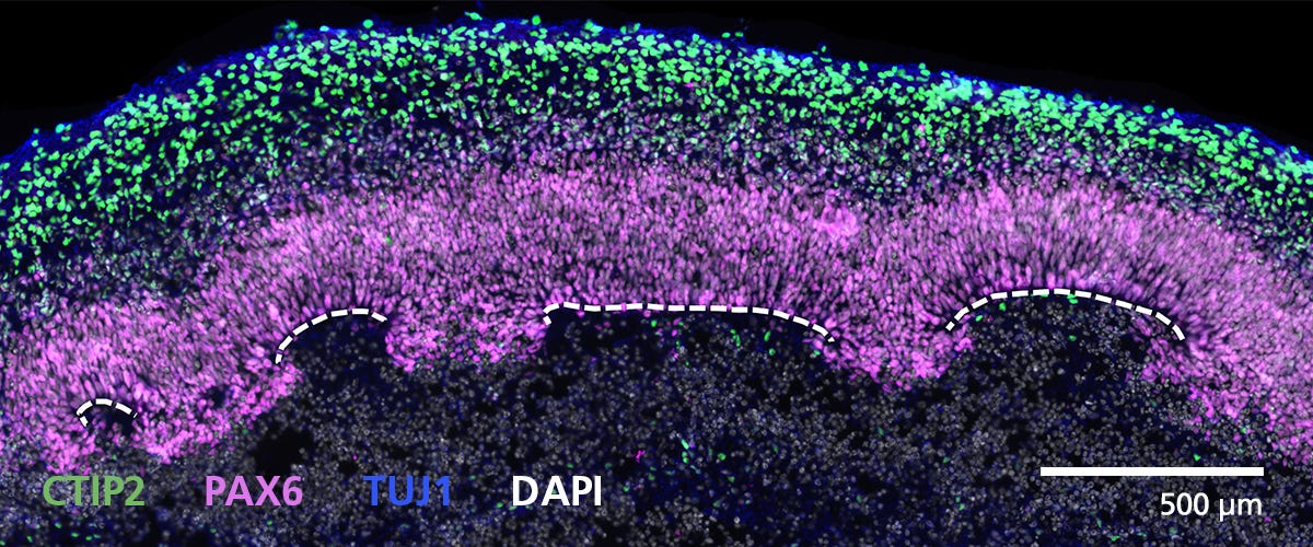
Figure 3. Cryosectioned Cerebral Organoids Show Stratification of Cortical Plate Neurons and Progenitor Zones

Cerebral organoids were generated using the STEMdiff™ Cerebral Organoid Kit. A 16-μm-thick section of a Day 40 cerebral organoid was stained for CTIP2 (green), PAX6 (magenta), βIII-tubulin/TUJ1 (blue), and DAPI (gray). Cortical regions are defined by progenitor cells (PAX6+) that are radially organized around a pseudo-ventricle (dashed line). These progenitors give rise to cortical plate neurons indicated by CTIP2 and TUJ1 expression. For a detailed cryogenic tissue processing and immunofluorescence protocol, please see the Methods Library.

Zones of Active Proliferation in Cerebral Organoids Are Preserved Following the Protocol for Tissue Processing

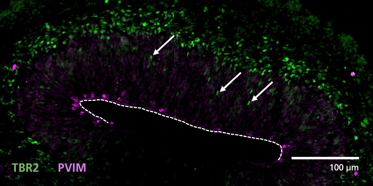
Figure 4. Zones of Active Proliferation in Cerebral Organoids Are Preserved Following the Protocol for Tissue Processing

Organoid tissue was processed for immunofluorescence and stained for TBR2 (intermediate precursors, green) and phosphorylated vimentin (PVIM, dividing cells, magenta). Cells actively divide at the apical border of cortical regions along the border of the pseudo-ventricle (dashed line). A population of these dividing cells will express TBR2 and then migrate (arrows) from the progenitor zone to form a layer of intermediate progenitors. For a detailed cryogenic tissue processing and immunofluorescence protocol, please see the Methods Library.

Immunofluorescence from Cryosectioned Cerebral Organoids Indicates Preserved Organization of Cortical Neurons

Figure 5. Immunofluorescence from Cryosectioned Cerebral Organoids Indicates Preserved Organization of Cortical Neurons

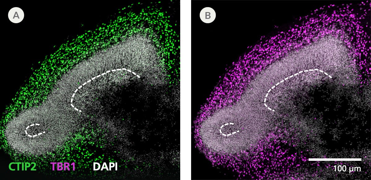
Organoid tissue was processed for immunofluorescence and stained for CTIP2 (green), TBR1 (layer 5/6 cortical neurons, magenta), and DAPI (white). Deep layer neuronal markers CTIP2 and TBR1 are expressed in cells around presumptive progenitor zones (dashed line) toward the outside or apical surface of organoids. For a detailed cryogenic tissue processing and immunofluorescence protocol, please see the Methods Library.

Cryogenic Tissue Processing and Immunofluorescence Captures Arrangement of Neural Progenitors Around Pseudo-Ventricles in Cerebral Organoids

Figure 6. Cryogenic Tissue Processing and Immunofluorescence Captures Arrangement of Neural Progenitors Around Pseudo-Ventricles in Cerebral Organoids

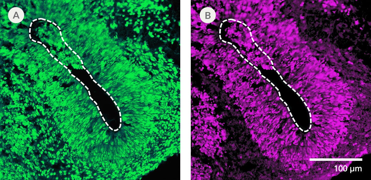
Organoid tissue was processed for immunofluorescence and stained for (A) FOXG1 (forebrain cells, green) or (B) SOX2 (neural progenitors, magenta). Organoids derived from STEMdiff™ Cerebral Organoid Kit generate forebrain-type tissue as indicated by FOXG1 expression. Neural progenitors expressing SOX2 are radially arranged around a pseudo-ventricle area (dashed line). For a detailed cryogenic tissue processing and immunofluorescence protocol, please see the Methods Library.

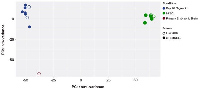
Cerebral Organoids Generated with the STEMdiff™ Cerebral Organoid Kit Are Transcriptionally Similar to Those from Published Protocols

Figure 7. Cerebral Organoids Generated with the STEMdiff™ Cerebral Organoid Kit Are Transcriptionally Similar to Those from Published Protocols

Principal component analysis of hPSC and cerebral organoid transcriptomes. Cerebral organoids generated using the STEMdiff™ Cerebral Organoid Kit (filled blue circles) cluster together, and cluster with previously published (C Luo et al. Cell Rep, 2016) cerebral organoids (open blue circles). The first principal component accounts for the majority of variance seen (PC1; 80%) and distinguishes the cerebral organoid samples from the hPSCs (green circles). The second principal component accounts for only 9% of the variation, and highlights the modest expression differences between cultured organoids and primary embryonic fetal brain samples (19 post-conceptional weeks, brown circles).

Neural Organoids Generated with STEMdiff™ Cerebral Organoid Kit Express Expected Key Markers

Figure 8. Neural Organoids Generated with STEMdiff™ Cerebral Organoid Kit Express Expected Key Markers

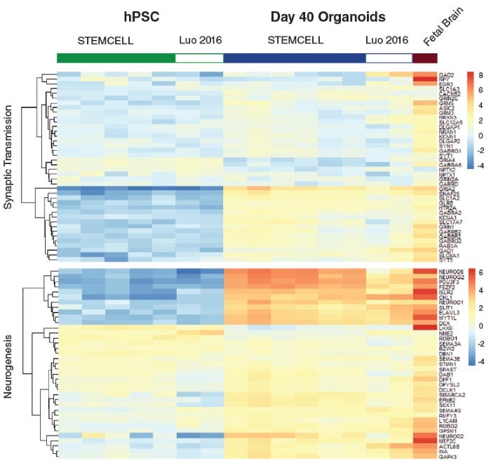
Heatmap of expression levels for genes associated with synaptic transmission function and neurogenesis in Day 40 organoids. These data show that gene expression of cerebral organoids generated from the STEMdiff™ Cerebral Organoid Kit are similar to published results (C Luo et al. Cell Rep, 2016).

scRNA seq data for STEMdiff™ Cerebral Organoid Kit

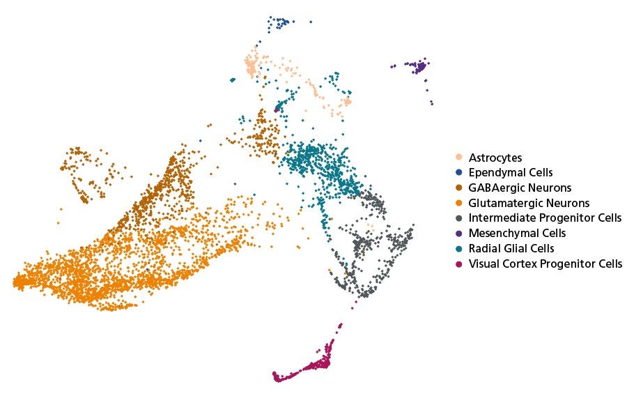
Figure 9. Neural Organoids Generated with STEMdiff™ Cerebral Organoid Kit Contain Diverse Cell Types

Unguided cerebral organoids were generated from H9 ESCs using STEMdiff™ Cerebral Organoid Kit. scRNA-seq gene expression data captures the cellular diversity of these neural organoids which can be further explored using the Single Cell RNA Sequencing Data Visualization Tool for Neural Organoids . At Day 50, the organoids were dissociated into a single-cell suspension. The library was prepared using Chromium Single Cell 3ʹ v1 protocol with Feature Barcoding technology (10x Genomics) following surface protein staining with TotalSeq™–B (BioLegend). The barcoded processing, gene counting, and aggregation were done using the Cell Ranger software v3.1.0. Further processing and demultiplexing was done with Seurat v4.1.1. The data have been made publicly available on GEO: GSE218457. ESC = embryonic stem cell

Time-lapse images and heatmap showing calcium signals spreading across a Day 91 cerebral organoid, indicating synchronized neuronal activity.

Figure 10. STEMdiff™ Cerebral Organoids Exhibit Synchronous Calcium Activity

Cerebral organoids were generated using the STEMdiff™ Cerebral Organoid Kit and matured with the STEMdiff™ Cerebral Organoid Maturation Kit. On Day 91, organoids were imaged using the ImageXpress® Confocal HT.ai High-Content Imaging System (Molecular Devices) with MetaXpress® High-Content Image Acquisition and Analysis Software. (A) Time-lapse images of a representative region within a cerebral organoid. Arrows indicate areas of elevated calcium signal intensity. (B) Corresponding heatmap representation of calcium intensity from the regions shown in (A) over time. Signal propagation from the initial peak to surrounding areas suggests the presence of a functional neuronal network.

Protocols and Documentation

Find supporting information and directions for use in the Product Information Sheet or explore additional protocols below.

Document Type
Product Name
Catalog #
Lot #
Language
Catalog #
08570
Lot #
All
Language
English
Document Type
Safety Data Sheet 1
Catalog #
08570
Lot #
All
Language
English
Document Type
Safety Data Sheet 2
Catalog #
08570
Lot #
All
Language
English
Document Type
Safety Data Sheet 3
Catalog #
08570
Lot #
All
Language
English
Document Type
Safety Data Sheet 4
Catalog #
08570
Lot #
All
Language
English
Document Type
Safety Data Sheet 5
Catalog #
08570
Lot #
All
Language
English
Document Type
Safety Data Sheet 6
Catalog #
08570
Lot #
All
Language
English
Document Type
Safety Data Sheet 7
Catalog #
08570
Lot #
All
Language
English

Applications

This product is designed for use in the following research area(s) as part of the highlighted workflow stage(s). Explore these workflows to learn more about the other products we offer to support each research area.

Resources and Publications

Educational Materials (48)

Publications (39)

Modeling Synaptic Maturation From Growth Cone to Synapse in Human Organoids M. S. Øhlenschlæger et al. Journal of Neurochemistry 2026 May

Abstract

Human neural organoids (NOs) provide a powerful platform for investigating synaptic development and dysfunction during early neurodevelopment. However, methodologies for isolating functional synaptic structures from these models remain limited. Here, we present a differential centrifugation protocol enabling the enrichment of growth cone particles (GCPs) and immature synaptosomes from air‐liquid interface cerebral organoids (ALI‐COs) at distinct developmental stages (Day 90 and 150). Notably, the method avoids density gradients, requires minimal starting material while maintaining reproducibility across human and murine tissues. Quantitative proteomic profiling revealed significant enrichment of growth cone markers (e.g., GAP43) and classical synaptosomal proteins (e.g., PCLO, BSN, SYN1). Transmission electron microscopy (TEM) confirmed the presence of membrane‐enclosed GCPs with fibrous content and mitochondria in Day 90 isolates, and immature synaptosomes containing synaptic vesicles on day 150. Functional viability of both types of synaptic structures was demonstrated through KCl‐induced depolarization, which triggered phosphorylation changes in growth cone proteins (GAP43, MARCKS, MARCKSL1), cytoskeletal regulators (DCLK1, SHTN1, MARK4, MAP1B) and protein kinases (CAMK2G, PRKCE) in Day 90 GCPs, as well as classical synaptic vesicle cycle proteins (SYN1, DNM1, RPH3A) at Day 150. Overall, this study establishes a centrifugation‐based protocol for isolating growth cones and immature synapses from human organoids, capturing key stages of synaptic development and enabling scalable, patient‐compatible models to study synaptic function and dysfunction in neurodevelopmental and neurodegenerative disorders. Synapses are implicated in several neurological disorders and psychiatric diseases. The emergence and wide use of neural organoids provide a new opportunity to study human synapses in healthy and disease settings. Therefore, we developed a simple method for the enrichment of synaptosomes and growth cone particles from forebrain organoids. The method is based on differential centrifugation, works with small tissue amounts, and is highly reproducible. We validated the functionality of the isolated structures using KCl stimulation and phosphoproteomics. The method enables detailed mapping of protein composition and function during growth cone pathfinding, synaptogenesis, and establishment of neural circuits in organoids.
Lysophagy protects against ANXA11 amyloid fibril toxicity and propagation in FTLD. H. Zheng et al. Translational neurodegeneration 2026 Jun

Abstract

BACKGROUND: Accumulation of Annexin A11 (ANXA11) aggregates is a distinct pathological hallmark of amyotrophic lateral sclerosis (ALS) and frontotemporal lobar degeneration (FTLD). While genetic studies have linked ANXA11 mutations (e.g., D40G) to disease, the precise molecular events converting aggregation into neurotoxicity and intercellular propagation remain elusive. We hypothesize that lysosomal integrity serves as a critical checkpoint in ANXA11 proteinopathy and that its failure drives disease progression. METHODS: To model the human pathology of ANXA11, we generated pre-formed fibrils (PFFs) of wild-type and FTLD/ALS-linked D40G mutant ANXA11. Human iPSC-derived neurons, 3D cerebral organoids, and bulk RNA-sequencing were employed to investigate neurotoxicity. High-resolution imaging, lentiviral knockdown, and biochemical assays were performed to delineate the lysosomal damage response and the subsequent "prion-like" spreading of aggregates. RESULTS: The internalized ANXA11 fibrils accumulated in lysosomes, triggering lysosomal membrane permeabilization (LMP). The D40G mutation exacerbated this toxicity, leading to severe LMP, mitochondrial depolarization, and specific transcriptional downregulation of the dynactin subunit ACTR10. Mechanistically, we identified a protective signaling axis involving p38 MAPK, MK2, and HSP27 that senses ANXA11-induced lysosomal damage and initiates lysophagy. Notably, in human cerebral organoids, failure of this lysophagic clearance facilitated the cytoplasmic escape of ANXA11, thereby accelerating its seeding activity and propagation to neighboring cells. Pharmacological or genetic modulation of this pathway significantly altered neuronal survival. CONCLUSIONS: Our study established lysosomal rupture as a primary driver of ANXA11-associated neurodegeneration and validated the p38/MK2/HSP27 axis as a crucial defense mechanism in human neural tissue. These findings provide a novel mechanistic link between lysosomal quality control and ANXA11 propagation, highlighting that enhancing lysophagic flux represents a promising translational strategy to halt the progression of FTLD and ALS.
CASPR2 Autoimmune Antibodies Induce Neuronal Hyperactivity in Human Brain Organoids A. R. Oliveira et al. Journal of Neurochemistry 2026 Feb

Abstract

Gestational transfer of brain‐reactive antibodies is a risk factor for neurodevelopmental disorders. Contactin‐associated protein‐like 2 (CASPR2) is a known target for pathogenic maternal autoantibodies which have been proposed to interfere with fetal neurodevelopment. However, the impact of CASPR2 antibodies on human brain development remains largely unknown. Here, to better understand the neurophysiological changes that occur in the presence of these pathogenic autoantibodies, we cultured unguided human neural organoids for a period of 6‐months in media containing anti‐CASPR2 antibodies. We then performed neurophysiological characterization via whole‐cell patch‐clamp and calcium imaging in acute organoid slices. Our results reveal that CASPR2 antibody exposure increased spontaneous synaptic activity, enhanced the maximal frequency of action potential firing and of spontaneous network activity. These findings are consistent with a state of neuronal hyperexcitability, a phenotype which is observed in several models of neurodevelopmental disorders. Mechanistically, the alterations observed in action potential waveform are in accordance with a role for CASPR2 in the regulation of voltage‐gated potassium channels and a pathological role for CASPR2 autoantibodies in driving neuronal hyperexcitability. Maternal antibodies targeting CASPR2 are a known risk factor for neurodevelopmental disorders, yet their impact on early human brain development remains unclear. We modeled this exposure using human neural organoids treated with patient‐derived CASPR2 antibodies up to the age of 6 months. Our study reveals that these antibodies drive neurons into a state of pathological hyperexcitability by specifically impairing action potential repolarization and enhancing excitatory synaptic transmission. These findings provide novel mechanistic evidence linking maternal autoimmunity to the excitation/inhibition imbalance characteristic of autism, highlighting a potential biological origin for antibody‐mediated neurodevelopmental conditions.
Need a high-quality cell source? Choose from our hiPSC healthy control lines, manufactured with mTeSR™ Plus.