ACCUTASE™

Cell detachment solution

ACCUTASE™

Cell detachment solution
From: 62 USD
Catalog #
(Select a product)
Cell detachment solution
Add to Wish List

Product Advantages


  • Obtain cleaner cultures with a ready-to-use solution, free from mammalian and bacterial-derived products

  • Achieve efficient cell detachment with this gentle trypsin alternative

  • Ensure high cell viability, without the need for a neutralizing solution

Overview

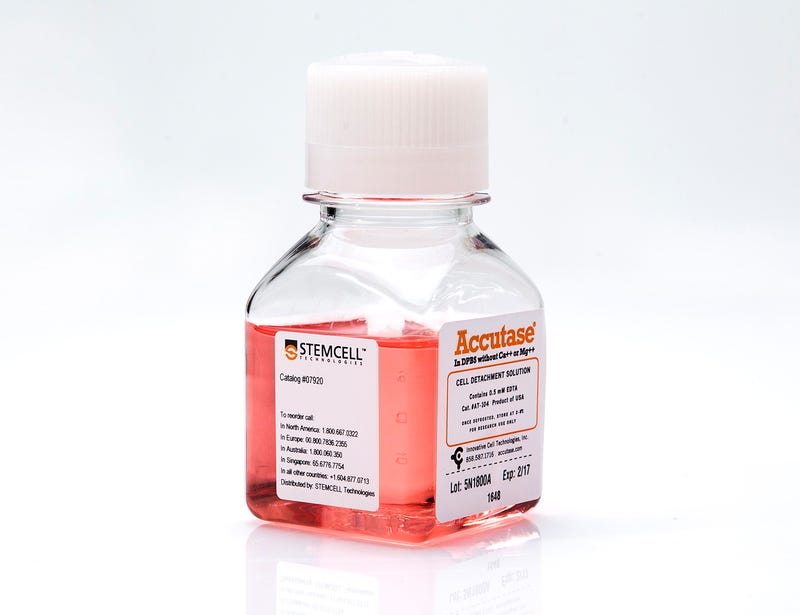
Use ACCUTASE™ for routine cell detachment from plasticware. This solution of proteolytic and collagenolytic enzymes comes ready-to-use and has been shown to work effectively across a wide variety of cell types. Formulated at a lower concentration than conventional trypsin, this gentle alternative preserves cell surface epitopes for downstream flow cytometry analyses and ensures high cell viability, without the need for a neutralizing solution. ACCUTASE™ does not contain mammalian or bacterial-derived products. Each lot of ACCUTASE™ is tested for sterility (by USP membrane filtration method), enzymatic activity (tested with synthetic chromagenic tetrapeptides), and cell detachment from tissue culture plastic.
Contains
• 1X ACCUTASE™ enzymes in Dulbecco’s phosphate-buffered saline (PBS)
• 0.5 mM EDTA•4Na
• 3 mg/L Phenol red
Subtype
Enzymatic
Cell Type
Neural Cells, PSC-Derived, Pluripotent Stem Cells
Species
Human, Mouse, Non-Human Primate, Other, Rat
Application
Cell Culture
Area of Interest
Neuroscience, Stem Cell Biology

Protocols and Documentation

Find supporting information and directions for use in the Product Information Sheet or explore additional protocols below.

Document Type
Product Name
Catalog #
Lot #
Language
Product Name
ACCUTASE™
Catalog #
07922, 07920
Lot #
All
Language
English
Document Type
Safety Data Sheet
Product Name
ACCUTASE™
Catalog #
07922, 07920
Lot #
All
Language
English

Resources and Publications

Publications (277)

CRISPR and Gene Augmentation Rescue Trabecular Meshwork Dysfunction in iPSC Models of Lowe Syndrome. S. Chen et al. Advanced science (Weinheim, Baden-Wurttemberg, Germany) 2026 Sep

Abstract

Lowe syndrome is a rare, currently incurable multisystem disorder that affects the eyes, kidneys, and central nervous system. It is caused by mutations in the OCRL gene, which encodes an inositol 5-phosphatase. The disorder remains incurable, and the pathways underlying the ocular symptoms remain poorly understood, largely due to the lack of appropriate models. In this study, trabecular meshwork cell models of Lowe syndrome were generated to test two distinct gene therapy strategies: a mutation-agnostic OCRL DNA augmentation therapy and a patient-specific CRISPR-mediated gene correction strategy. The results showed that AAV2-OCRL demonstrated the highest transduction efficiency in patient iPSC-derived trabecular meshwork models (iHTM) among the three AAV-OCRL vectors evaluated, establishing it as a promising delivery vector. Targeted CRISPR-based gene therapy restored OCRL enzyme activity and corrected cellular defects in patient iPSC-derived trabecular meshwork cell models. Furthermore, RNA-sequencing analysis of these models revealed dysregulation of extracellular matrix organization, cell adhesion, focal adhesion, and cytoskeletal regulatory pathways, suggesting that disruption of interconnected ECM-adhesion-cytoskeletal networks may contribute to trabecular meshwork dysfunction in Lowe syndrome-associated glaucoma. These findings indicate the feasibility of OCRL gene augmentation and CRISPR-based gene editing in patient-derived ocular models and position AAV2-OCRL as a leading therapeutic candidate for Lowe syndrome.
Clonal LIMD1 loss drives PD-L1 immune evasion via ARIH1-dependent ubiquitination in lung cancer. K. Shah et al. Life science alliance 2026 Oct

Abstract

LIMD1, a tumour suppressor located at chromosome 3p21.3, is frequently lost in non-small-cell lung cancer, yet its role in tumour-immune interactions remains unclear. Here, we show LIMD1 loss increases PD-L1 protein abundance across multiple lung cancer models and primary airway epithelial cells. Mechanistically, LIMD1 restrains PD-L1 through post-transcriptional and post-translational mechanisms. LIMD1 loss can relieve microRNA-mediated repression of the CD274 3'UTR, and LIMD1 loss can also disrupt ARIH1-PD-L1 association, reduce PD-L1 polyubiquitination, and stabilise PD-L1 protein without a commensurate increase in CD274 transcript levels in isogenic models. Functionally, LIMD1-deficient tumour cells suppress CD8+ T-cell activation in vitro and show enhanced sensitivity to PD-1/PD-L1 blockade in tumour-PBMC co-culture assays. Analysis of TRACERx non-small-cell lung cancer samples revealed clonal LIMD1 loss of heterozygosity in ∼40% of lung adenocarcinomas, where it is associated with increased tumour PD-L1 expression. Across independent patient cohorts receiving immune checkpoint blockade, low LIMD1 expression was enriched among responders. We identify LIMD1 as a tumour-intrinsic regulator of PD-L1 turnover and suggest that tumour suppressor loss can shape immune checkpoint biology and influence immunotherapy response.
Generation of Functional Patient-Specific Thymus Organoids From Human Pluripotent Stem Cells (hPSCs) Using Air–Liquid Interface Culture S. A. Ramos, H. A. Russ Bio-protocol 2026 May

Abstract

The thymus is critical for the establishment of a functional and self-tolerant adaptive immune system, but it involutes with age, resulting in reduced naive T-cell output. Generation of a functional human thymus from human pluripotent stem cells (hPSCs) is an attractive regenerative medicine strategy. Direct differentiation of thymic epithelial progenitors (TEPs) from hPSCs has been demonstrated in vitro, but functional thymic epithelial cells (TECs) develop only after transplantation of TEPs in vivo. Functional human reaggregated thymic organoid cultures (RTOCs) and artificial thymic organoids (ATOs) cultured at the air–liquid interface support T-cell development in vitro and in vivo and permit the interrogation of human thymic function and T-cell development. However, these approaches require access to primary human tissues or murine bone marrow stromal cells, are allogeneic, and do not support negative selection. Recently, we reported the directed differentiation of induced PSCs (iPSCs) to functional thymic epithelial progenitors (TEPs) that support murine T-cell development after transplantation in nude mice. Here, we combined hPSC-derived TEPs, hematopoietic progenitor cells (HPCs), and mesenchymal cells, differentiated from the same hPSC line, and generated functional isogenic stem cell–derived thymic organoids (sTOs). Our revised protocol improves our TEP differentiation process and allows the generation of functional isogenic, patient-specific thymic organoids in vitro.