hPSC Genetic Analysis Kit

qPCR analysis kit for detecting the majority of karyotypic abnormalities reported in human ESCs and iPSCs
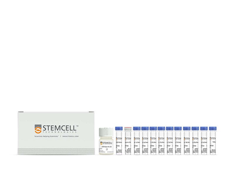
hPSC Genetic Analysis Kit

qPCR analysis kit for detecting the majority of karyotypic abnormalities reported in human ESCs and iPSCs

From: 819 USD
Catalog #
(Select a product)
qPCR analysis kit for detecting the majority of karyotypic abnormalities reported in human ESCs and iPSCs
Add to Wish List

What's Included

  • qPCR Master Mix (2X), 3 mL
  • ROX Reference Dye, 0.2 mL
  • Chr 1q Genetic Assay, 60 Rxn
  • Chr 4p Genetic Assay, 60 Rxn
  • Chr 8q Genetic Assay, 60 Rxn
  • Chr 10p Genetic Assay, 60 Rxn
  • Chr 12p Genetic Assay, 60 Rxn
  • Chr 17q Genetic Assay, 60 Rxn
  • Chr 18q Genetic Assay, 60 Rxn
  • Chr 20q Genetic Assay, 60 Rxn
  • Chr Xp Genetic Assay, 60 Rxn
  • Genomic DNA Control, 15 µL
  • TE Buffer, 1 mL
Products for Your Protocol
To see all required products for your protocol, please consult the Protocols and Documentation.

Overview

Quickly and cost-effectively detect the 8 most common karyotypic abnormalities reported in human pluripotent stem cells (hPSCs).

This qPCR-based kit enables the genetic screening of multiple hPSC lines and contains enough material to analyze a maximum of 18 samples and 2 controls in triplicate including control DNA. It uses double-quenched probes with a 5-carboxyfluorescein (5-FAM) dye to give superior performance over other, single-quenched probes. The kit includes qPCR Master Mix, ROX Reference Dye, Buffer, and primer/probe mixes designed to detect the critical minimal regions of the 8 most commonly mutated regions, as well as a primer/probe mix to a control location in the genome. A genomic DNA control is also provided with demonstrated normal copy number over the regions of interest.

To understand your results, input qPCR data into our online Genetic Analysis App, which will perform statistical analyses, assist with data interpretation, and provide visual representation of the data
Cell Type
Pluripotent Stem Cells
Species
Human
Application
Genome Editing
Area of Interest
Disease Modeling, Stem Cell Biology

Data Figures

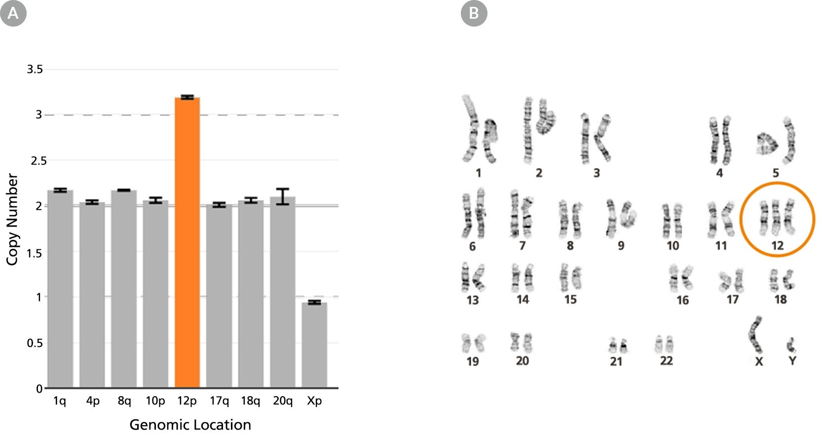
Figure 1. The hPSC Genetic Analysis Kit Identifies Chromosome 12 Trisomy

Chromosome 12 trisomy in WLS-1C human iPS cell line is (A) detected using the hPSC Genetic Analysis Kit (orange bar) and (B) confirmed by G-banding.

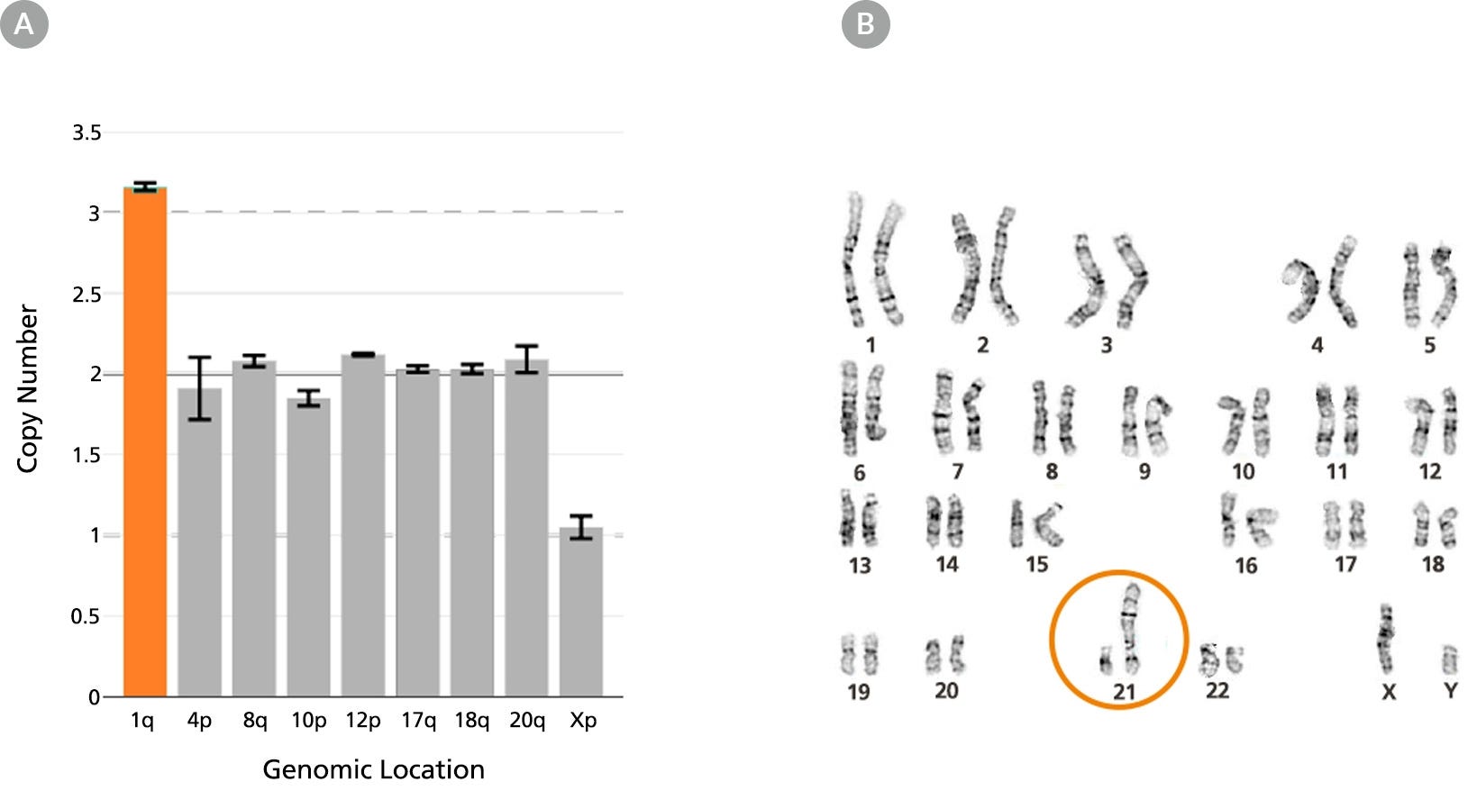
Figure 2. The hPSC Genetic Analysis Kit Identifies Chromosome 1 Duplication via Unbalanced Translocation

Unbalanced rearrangement of chromosome 1 in the WLS-1C human iPS cell line in which an extra copy of the long (q) arm of chromosome 1 translocated to the short arm (p) of chromosome 21 was (A) detected using the hPSC Genetic Analysis Kit (orange bar) and (B) confirmed by G-banding.

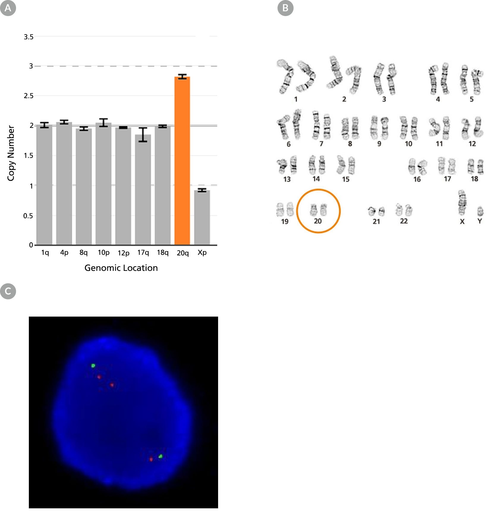
Figure 3. The hPSC Genetic Analysis Kit Identifies Chromosome 20q11.21 Duplication

Chromosome 20q duplication in WLS-1C human iPS cell line is (A) detected using the hPSC Genetic Analysis Kit (orange bar), (B) undetected by G-banding, and (C) confirmed by fluorescent in situ hybridization using probes for 20p11 (green) and 20q11.21 (red).

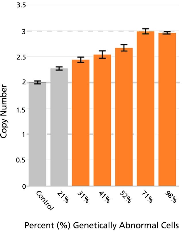
Figure 4. The hPSC Genetic Analysis Kit Identifies Abnormalities in Cultures with Approximately 30% Mosaicism

Genetically normal WLS-1C human iPS cells were mixed in the indicated ratios with WLS-1C human iPS cells containing a chromosome 20q duplication. Cultures with approximately 30% genetically abnormal cells exhibit a significantly enriched population of 20q11.21 duplication (orange bars).

Protocols and Documentation

Find supporting information and directions for use in the Product Information Sheet or explore additional protocols below.

Document Type
Product Name
Catalog #
Lot #
Language
Catalog #
07550
Lot #
All
Language
English
Document Type
Safety Data Sheet 1
Catalog #
07550
Lot #
All
Language
English
Document Type
Safety Data Sheet 2
Catalog #
07550
Lot #
All
Language
English
Document Type
Safety Data Sheet 3
Catalog #
07550
Lot #
All
Language
English
Document Type
Safety Data Sheet 4
Catalog #
07550
Lot #
All
Language
English
Document Type
Safety Data Sheet 5
Catalog #
07550
Lot #
All
Language
English
Document Type
Safety Data Sheet 6
Catalog #
07550
Lot #
All
Language
English
Document Type
Safety Data Sheet 7
Catalog #
07550
Lot #
All
Language
English
Document Type
Safety Data Sheet 8
Catalog #
07550
Lot #
All
Language
English
Document Type
Safety Data Sheet 9
Catalog #
07550
Lot #
All
Language
English
Catalog #
07550
Lot #
All
Language
English
Catalog #
07550
Lot #
All
Language
English
Catalog #
07550
Lot #
All
Language
English
Catalog #
07550
Lot #
All
Language
English

Applications

This product is designed for use in the following research area(s) as part of the highlighted workflow stage(s). Explore these workflows to learn more about the other products we offer to support each research area.

Resources and Publications

Educational Materials (38)

Publications (19)

De novo EHMT2 variants cause an autosomal dominant EHMT2-related Kleefstra syndrome via loss of G9a methyltransferase activity. A. Hnízda et al. Nature communications 2026 Jul

Abstract

EHMT1 and EHMT2 genes encode human euchromatin histone lysine methyltransferase 1 and 2 (EHMT1 alias GLP; EHMT2 alias G9a) that form heteromeric GLP/G9a complexes with essential roles in epigenetic regulation of gene expression. While EHMT1 haploinsufficiency has been established as the cause of Kleefstra syndrome 1, the pathogenesis of G9a dysfunction in human disease remains largely unknown. We identified seven de novo EHMT2 variants in patients with clinical presentation, episignatures, histone modifications and transcriptomic profiles similar to those of Kleefstra syndrome 1. In vitro studies reveal that these variants encode for structurally stable G9a proteins that are catalytically incompetent due to aberrant interactions either with histone H3 tail or with S-adenosylmethionine. Heterozygous mice carrying a patient-derived variant exhibit growth retardation, facial/skull dysmorphia and aberrant behavior. Here we report pathogenic EHMT2 variants that likely exert dominant-negative effect on GLP/G9a complexes and thus genocopy the EHMT1 haploinsufficiency via a distinct molecular mechanism, defining an autosomal dominant EHMT2-related Kleefstra syndrome.
Kat5 cKO mouse replicates biological domain signatures associated with Alzheimer's disease. G. Cary et al. Alzheimer's & dementia : the journal of the Alzheimer's Association 2026 Jul

Abstract

INTRODUCTION: Alzheimer's disease (AD) can be caused by autosomal-dominant familial Alzheimer's disease (FAD) mutations in amyloid precursor protein (APP) or presenilin-1 and 2, which form an enzyme substrate complex. KAT5 binds to the APP intracellular domain. Recent reports of decreased γ-secretase activity in FAD mutants support KAT5 membrane sequestration. METHODS: We compare the hippocampal transcriptome profiles of the Kat5 brain-specific knockout (KO) mouse to multiple AD datasets through alignment with the TREAT-AD AD biological domains. We examine KAT5 subcellular localization in human wild-type and AD neurons. RESULTS: The Kat5 KO mouse demonstrates downregulation of synaptic genes, metabolic pathways, and upregulation of DNA replication and repair, cell cycle, and immune response genes. We see similar profiles in Kat5 and comparative AD datasets. KAT5 is restricted to the cytosol in human AD neurons. DISCUSSION: This analysis supports the hypothesis that KAT5 nuclear signaling downstream of APP cleavage plays a pivotal role in neuronal homeostasis.
A stem cell-based toolkit to model Angelman syndrome caused by paternal uniparental disomy of chromosome 15 F. C. Mateus et al. Human Cell 2025 Sep

Abstract

Angelman syndrome is a rare neurodevelopmental disorder caused by the loss of function of the maternally inherited UBE3A gene within the chr15q11-q13 region. This gene is subjected to a tissue-specific form of genomic imprinting leading to the silencing of the paternal allele in neurons. Angelman syndrome can result from various (epi)genetic mechanisms, with paternal uniparental disomy of chromosome 15 (patUPD15) being one of the rarest and least studied due to the absence of suitable models. To address this gap, we generated three independent induced pluripotent stem cell (iPSC) lines from individuals with Angelman syndrome caused by patUPD15, alongside genetically matched unaffected familial controls. Peripheral blood mononuclear cells (PBMCs) were reprogrammed into iPSCs using a non-integrative Sendai virus-based approach expressing the Yamanaka factors. All iPSC lines underwent rigorous quality control, confirming stem cell identity, trilineage differentiation potential, and genetic and epigenetic integrity. This newly established iPSC toolkit provides a powerful platform to investigate the molecular underpinnings of Angelman syndrome caused by patUPD15, paving the way for future translational research and therapeutic development tailored for this understudied form of the disorder. The online version contains supplementary material available at 10.1007/s13577-025-01287-8.