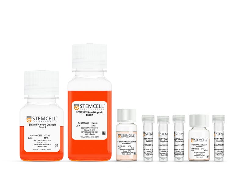
STEMdiff™ Choroid Plexus Organoid Differentiation Kit
Culture medium kit for establishment of organoid models containing human pluripotent stem cell-derived choroid plexus-like epithelium
Request Pricing
Thank you for your interest in this product. Please provide us with your contact information and your local representative will contact you with a customized quote. Where appropriate, they can also assist you with a(n):
Estimated delivery time for your area
Product sample or exclusive offer
In-lab demonstration
-
 Gentle Cell Dissociation Reagent
cGMP, enzyme-free cell dissociation reagent
-
 STEMdiff™ Choroid Plexus Organoid Maturatio...
Culture medium kit for establishment of organoid models containing human pluripotent stem cell-derived choroid plexus-like epithelium
-
 Ultra-Low Adherent Plate for Suspension Cultu...
Sterile, flat-bottom plate, with ultra-low adherent surface for suspension cultures, with lid; 6-well format
-
 Y-27632 (Dihydrochloride)
RHO/ROCK pathway inhibitor; Inhibits ROCK1 and ROCK2
-
Labeling Antibodies
Compatible antibodies for purity assessment of isolated cells
Overview
Using STEMdiff™ Choroid Plexus Differentiation Kit’s simple five-stage protocol, you can efficiently and reproducibly generate choroid plexus organoids for a variety of downstream applications. The kit is based on the formulation published by Pellegrini et al. (Science, 2020), which was awarded a 3Rs Prize for its breakthroughs in animal reduction, replacement, and refinement. Choroid plexus organoid formation is initiated through an intermediate embryoid body (EB) formation step followed by expansion of neuroepithelia and patterning to choroid plexus-like epithelium. After a maturation period, organoids generated using this kit feature cystic structures filled with a fluid resembling CSF and surrounded by an epithelial layer expressing ependymal choroid plexus-specific markers (TTR, CLIC6, AQP1).
For extended periods of organoid culture (> 40 days), the components required for organoid maturation can be purchased as STEMdiff™ Choroid Plexus Organoid Maturation Kit (Catalog #100-0825).
Data Figures
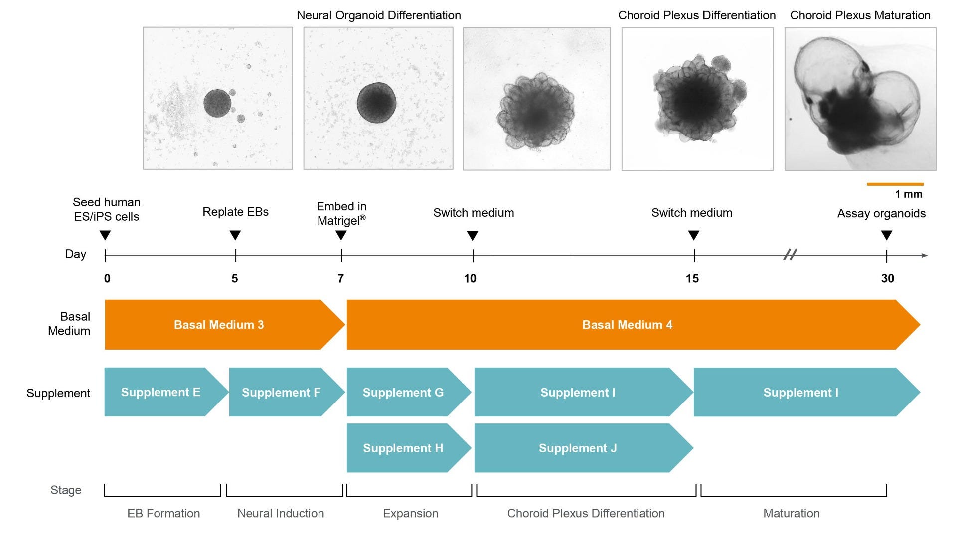
Figure 1. Protocol Schematic for the STEMdiff™ Choroid Plexus Organoid Differentiation and Maturation Kits
Choroid plexus organoids can be generated from human embryonic stem (ES) or induced pluripotent stem (iPS) cells in 30 days. The protocol begins with embryoid body (EB) formation, followed by expansion of neuroepithelia and patterning to choroid plexus-like epithelium. After a period of epithelial maturation including extensive bubbling, the organoids develop cystic structures surrounded by an ependymal epithelial layer and filled with a fluid resembling cerebrospinal fluid (CSF).
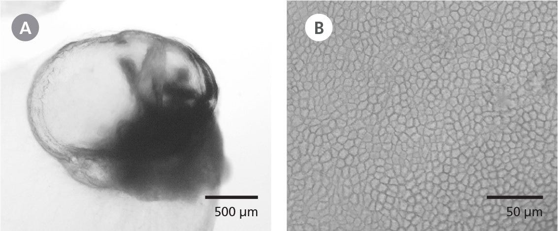
Figure 2. Organoids Generated Using the STEMdiff™ Choroid Plexus Organoid Differentiation and Maturation Kits Exhibit Fluid-Filled Cysts and Ependymal Epithelial Morphology
(A) Phase-contrast image of a whole choroid plexus organoid at day 40 using the STEMdiff™ Choroid Plexus Organoid Differentiation and Maturation Kits. Organoids exhibit translucent cysts containing a CSF-like fluid. (B) Epithelia on choroid plexus organoids exhibit a tightly packed and cuboidal morphology typical of ependymal cells that line the brain ventricles in vivo.
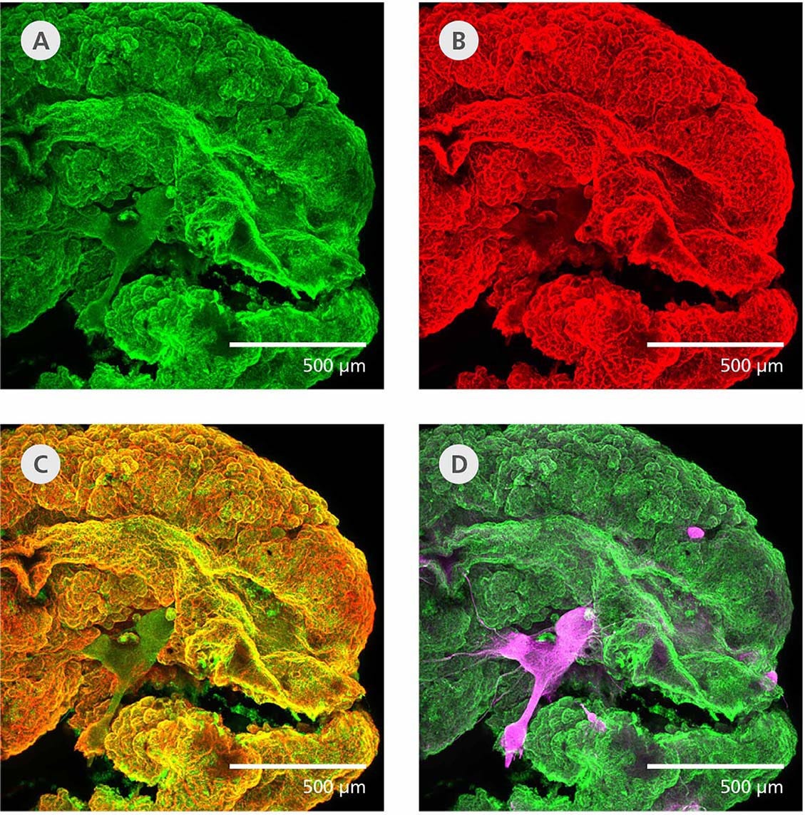
Figure 3. Choroid Plexus Organoids Express Markers of Ependymal Epithelium and Show Limited Expression of Neuroepithelial Markers
Immunohistological analysis on whole cleared and stained (Masselink et al. 2019) choroid plexus organoids at day 40 reveals that their cystic structures are (A) TTR- (green) and (B) CLIC6- (red) positive. (C) These ependymal cell markers are highly co-localized. (D) Bundles of MAP2-positive (magenta) neurons are observed but are not co-localized with the ependymal cell markers.
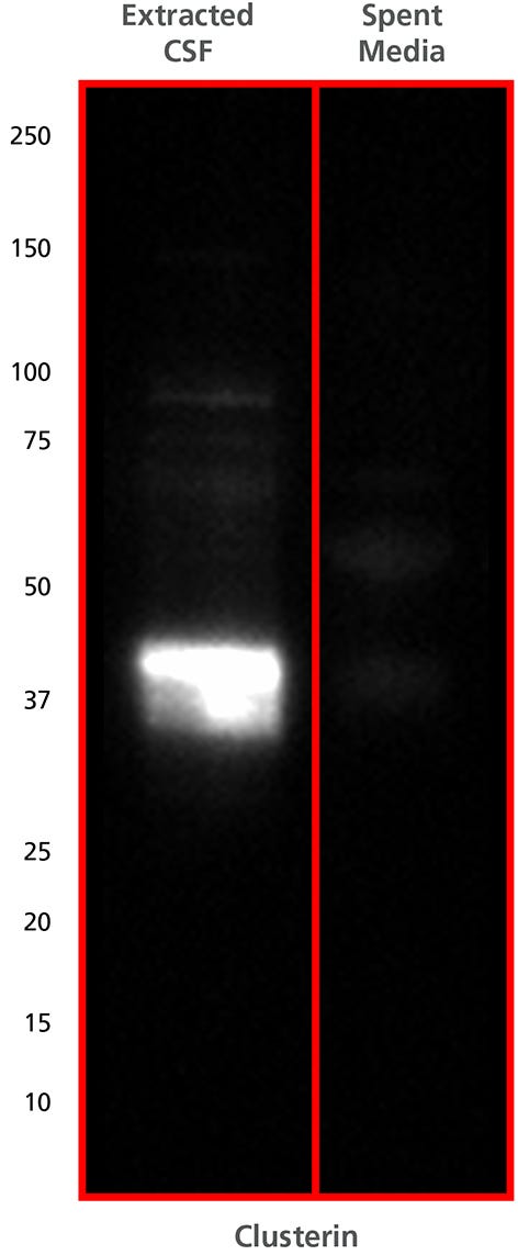
Figure 4. Fluid Extracted from Cysts in Choroid Plexus Organoids is Enriched with Clusterin Protein, A Marker of Cerebrospinal Fluid (CSF)
CSF-like fluid was extracted from cysts contained in day 40 choroid plexus organoids using a 28G syringe. A western blot was performed on the extracted fluid to detect Clusterin and shows a band between the 37 and 50 kDa molecular weight marker. Clusterin is a soluble secreted chaperone protein found in high abundance in CSF.
Protocols and Documentation
Find supporting information and directions for use in the Product Information Sheet or explore additional protocols below.
Applications
This product is designed for use in the following research area(s) as part of the highlighted workflow stage(s). Explore these workflows to learn more about the other products we offer to support each research area.
Resources and Publications
Educational Materials (13)
Related Products
-
 STEMdiff™ Cerebral Organoid Kit
Culture medium kit for establishment and maturation of human cerebral organoids
-
 Organoid Embedding Sheet
Silicone sheet designed to facilitate embedding 3D aggregates in a matrix
-
 STEMdiff™ Dorsal Forebrain Organoid Differe...
Cell culture medium kit for robust generation of dorsal forebrain neural organoids from human pluripotent stem cells
-
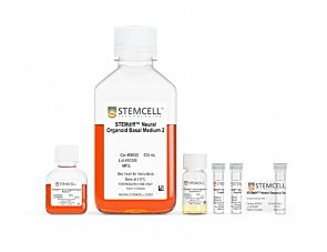 STEMdiff™ Ventral Forebrain Organoid Differ...
Cell culture medium kit for robust generation of ventral forebrain neural organoids from human pluripotent stem cells
Item added to your cart

STEMdiff™ Choroid Plexus Organoid Differentiation Kit
Quality Statement:
PRODUCTS ARE FOR RESEARCH USE ONLY AND NOT INTENDED FOR HUMAN OR ANIMAL DIAGNOSTIC OR THERAPEUTIC USES UNLESS OTHERWISE STATED. FOR ADDITIONAL INFORMATION ON QUALITY AT STEMCELL, REFER TO WWW.STEMCELL.COM/COMPLIANCE.
