Make more informed purchasing decisions with our new product availability and delivery estimate feature, now available on all product pages, in your cart, and during checkout.
Sign In
New to STEMCELL?
Register for an account to quickly and easily purchase products online and for one-click access to all educational content.
Thank you for your interest in this product.
Please provide us with your contact information and your local representative
will contact you with a customized quote. Where appropriate, they can also assist you with a(n):
Estimated delivery time for your area
Product sample or exclusive offer
In-lab demonstration
By submitting this form, you are providing your consent to STEMCELL Technologies Canada Inc. and its subsidiaries and affiliates (“STEMCELL”) to collect and use your information, and send you newsletters and emails in accordance with our privacy policy. Please contact us with any questions that you may have. You can unsubscribe or change your email preferences at any time.
Support the growth and differentiation of human embryonic stem (ES) and induced pluripotent stem (iPS) cells under feeder-free conditions with this defined and xeno-free cell culture matrix.
For consistent cell populations and reproducible results in downstream applications, use CellAdhere™ Laminin-521 with TeSR™ maintenance media to provide a defined culture substrate for cell maintenance. Laminin 521 is expressed and secreted by human pluripotent stem cells (hPSCs) in the inner cell mass of the embryo and therefore creates a biologically relevant hPSC culture environment in vitro. Use CellAdhere™ Laminin-521 with eTeSR™ (Catalog #100-1215) maintenance medium for single-cell passaging. Compared to other matrices, CellAdhere™ Laminin-521 increases single-cell attachment and survival and does not require the addition of apoptotic inhibitors during long term culture. Pair with Gentle Cell Dissociation Reagent (GCDR; Catalog #07174) or ReLeSR™ (Catalog #05872) for routine passaging of PSC aggregates, or Accutase™ (Catalog #07920) for single-cell passaging workflows.
Note that single-cell passaging of human ES and iPS cells can result in selective pressure and lead to genetic aberrations. If passaging as single cells, we recommend checking the karyotype frequently.
This product is designed for use in the following research area(s) as part
of the highlighted workflow stage(s). Explore these workflows to learn more about the other products we
offer to support each research area.
Rapid generation of purified human RPE from pluripotent stem cells using 2D cultures and lipoprotein uptake-based sorting
F. Michelet et al.
Stem cell research {\&} therapy 2020
Abstract
BACKGROUND: Despite increasing demand, current protocols for human pluripotent stem cell (hPSC)-derived retinal pigment epithelium (RPE) remain time, labor, and cost intensive. Additionally, absence of robust methods for selective RPE purification and removal of non-RPE cell impurities prevents upscaling of clinical quality RPE production. We aimed to address these challenges by developing a simplified hPSC-derived RPE production and purification system that yields high-quality RPE monolayers within 90 days. METHODS: Human pluripotent stem cells were differentiated into RPE using an innovative time and cost-effective protocol relying entirely on 2D cultures and minimal use of cytokines. Once RPE identity was obtained, cells were transferred onto permeable membranes to acquire mature RPE morphology. RPE differentiation was verified by electron microscopy, polarized VEGF expression, establishment of high transepithelial electrical resistance and photoreceptor phagocytosis assay. After 4 weeks on permeable membranes, RPE cell cultures were incubated with Dil-AcLDL (DiI-conjugated acetylated low-density lipoproteins) and subjected to fluorescence-activated cell sorting (FACS) for purification and subculture. RESULTS: Using our 2D cytokine scarce protocol, hPSC-derived functional RPE cells can be obtained within 2 months. Nevertheless, at this stage, most samples contain a percentage of non-RPE/early RPE progenitor cells that make them unsuitable for clinical application. We demonstrate that functional RPE cells express high levels of lipoprotein receptors and that this correlates with their ability to uptake lipoproteins. Combining photoreceptor uptake assay with lipoprotein uptake assay further confirms that only functional RPE cells uptake AcLDL. Incubation of mixed RPE/non-RPE cell cultures with fluorophore conjugated AcLDL and subsequent FACS-based isolation of labeled cells allows selective purification of mature functional RPE. When subcultured, DiI-AcLDL-labeled cells rapidly form pure homogenous high-quality RPE monolayers. CONCLUSIONS: Pure functional RPE monolayers can be derived from hPSC within 90 days using simplified 2D cultures in conjunction with our RPE PLUS protocol (RPE Purification by Lipoprotein Uptake-based Sorting). The simplicity of this protocol makes it scalable, and the rapidity of production and purification allows for high-quality RPE to be produced in a short span of time making them ideally suited for downstream clinical and in vitro applications.
WNT inhibition and increased FGF signaling promotes derivation of less heterogeneous primed human embryonic stem cells, compatible with differentiation
J. Taelman et al.
Stem Cells and Development 2019
Abstract
Human embryonic stem cells (hESCs) hold great value for future clinical applications. However, standard culture conditions maintain hESCs in a primed state, which bears heterogeneity in pluripotency and a tendency for spontaneous differentiation. To counter these drawbacks, primed hESCs have been converted to a naive state, but this has restricted the efficiency of existing directed differentiation protocols. In mouse, WNT inhibition by inhibitor of WNT production-2, together with a higher dose of fibroblast growth factor 2 (12 ng/mL) in DMEM/F12 basal medium (DhiFI), markedly improved derivation and maintenance of primed mouse epiblast stem cells. In this study, we show that DhiFI conditions similarly improved primed hESC traits, such as conferring a primed transcriptional signature with high levels of pluripotency markers and reduced levels of differentiation markers. When triggered to differentiate to neuronal and cardiac lineages, DhiFI hESCs and isogenic primed hESCs progressed similarly. Moreover, DhiFI conditions supported the derivation of hESC lines from a post-inner cell mass intermediate (PICMI). DhiFI-derived hESCs showed less spontaneous differentiation and expressed significantly lower levels of lineage-specific markers, compared to primed-derived lines from the same PICMI. Overall, DhiFI hESCs retained advantages of both primed and naive pluripotency and may ultimately represent a more favorable starting point for differentiation toward clinically desired cell types.
Chromosome Segregation Fidelity in Epithelia Requires Tissue Architecture
K. A. Knouse et al.
Cell 2018
Abstract
Much of our understanding of chromosome segregation is based on cell culture systems. Here, we examine the importance of the tissue environment for chromosome segregation by comparing chromosome segregation fidelity across several primary cell types in native and nonnative contexts. We discover that epithelial cells have increased chromosome missegregation outside of their native tissues. Using organoid culture systems, we show that tissue architecture, specifically integrin function, is required for accurate chromosome segregation. We find that tissue architecture enhances the correction of merotelic microtubule-kinetochore attachments, and this is especially important for maintaining chromosome stability in the polyploid liver. We propose that disruption of tissue architecture could underlie the widespread chromosome instability across epithelial cancers. Moreover, our findings highlight the extent to which extracellular context can influence intrinsic cellular processes and the limitations of cell culture systems for studying cells that naturally function within a tissue. Tissue architecture and integrin function are critical factors that support chromosome segregation fidelity in epithelial tissues.
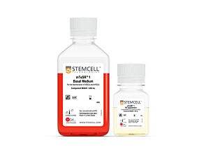
cGMP, feeder-free maintenance medium for human ES and iPS cells
Item added to your cart
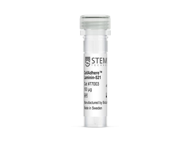
CellAdhere™ Laminin-521
Quality Statement:
PRODUCTS ARE FOR RESEARCH USE ONLY AND NOT INTENDED FOR HUMAN OR ANIMAL DIAGNOSTIC OR THERAPEUTIC USES UNLESS OTHERWISE STATED. FOR ADDITIONAL INFORMATION ON QUALITY AT STEMCELL, REFER TO WWW.STEMCELL.COM/COMPLIANCE.