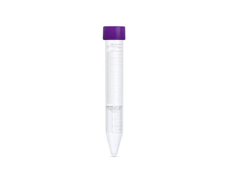
SepMate™-15 (IVD)
Tube for density gradient centrifugation for in vitro diagnostic (IVD) applications
Request Pricing
Thank you for your interest in this product. Please provide us with your contact information and your local representative will contact you with a customized quote. Where appropriate, they can also assist you with a(n):
Estimated delivery time for your area
Product sample or exclusive offer
In-lab demonstration
-
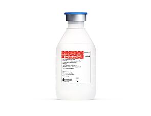 Lymphoprep™
Density gradient medium for the isolation of mononuclear cells
-
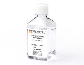 Dulbecco's Phosphate Buffered Saline with 2% ...
Cell culture buffer
What Our Scientist Says
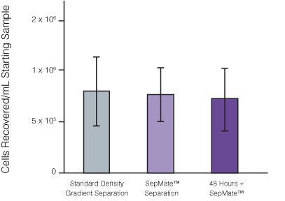
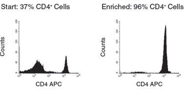
Traditional isolation of PBMCs requires careful layering of blood onto density gradient media prior to centrifugation. We developed SepMate™ to simplify this process, so anyone can isolate PBMCs with a simple pour while maintaining consistency across samples.

Overview
Data Figures
Protocols and Documentation
Find supporting information and directions for use in the Product Information Sheet or explore additional protocols below.
Applications
This product is designed for use in the following research area(s) as part of the highlighted workflow stage(s). Explore these workflows to learn more about the other products we offer to support each research area.
Resources and Publications
Educational Materials (13)


Publications (30)
Abstract
Abstract
Abstract
Related Products

SepMate™ (IVD) is only available in regions where it is registered as an In Vitro Diagnostic (IVD) device for the isolation of MNCs from whole blood or bone marrow by density gradient centrifugation. SepMate™ is manufactured under a cGMP quality managment system compliant to 21 CFR 820.
PRODUCTS ARE FOR RESEARCH USE ONLY AND NOT INTENDED FOR HUMAN OR ANIMAL DIAGNOSTIC OR THERAPEUTIC USES UNLESS OTHERWISE STATED. FOR ADDITIONAL INFORMATION ON QUALITY AT STEMCELL, REFER TO WWW.STEMCELL.COM/COMPLIANCE.
