
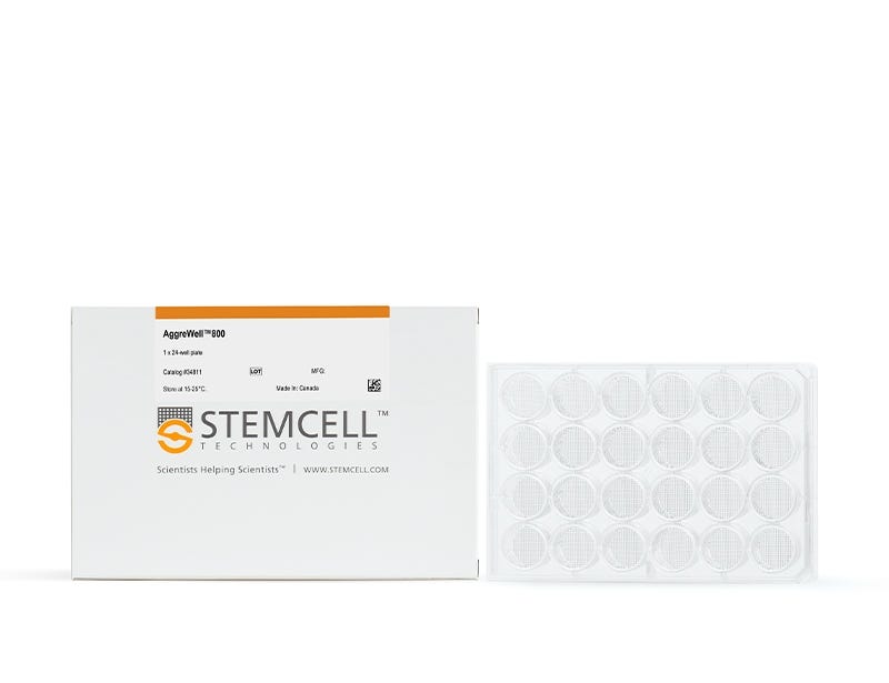
AggreWell™800
Microwell culture plates for easy and reproducible production of embryoid bodies and spheroids
Products for Your Protocol
To see all required products for your protocol, please consult the
Protocols and Documentation.
-
 Anti-Adherence Rinsing Solution
Rinsing solution for cultureware to prevent cell adhesion
Overview
Protocols and Documentation
Find supporting information and directions for use in the Product Information Sheet or explore additional protocols below.
Applications
This product is designed for use in the following research area(s) as part of the highlighted workflow stage(s). Explore these workflows to learn more about the other products we offer to support each research area.
Resources and Publications
Educational Materials (21)


Publications (64)
AHA1 Regulates Aβ Production via Modulation of APP Abundance and γ-Secretase Assembly.
Journal of neurochemistry 2026 Sep
Abstract
Deposition of amyloid β-protein (Aβ) is a hallmark of Alzheimer's disease (AD), produced by γ-secretase-mediated cleavage of amyloid precursor protein (APP). The 90-kDa heat shock protein (Hsp90) co-chaperone, activator of Hsp90 ATPase homolog 1 (AHA1), is known to promote the accumulation of toxic tau species; however, its effects on Aβ production remain unclear. Using cellular models-including human embryonic kidney 293 T (HEK293T) cells, stable HEK-APP cells, presenilin 1/2 double-knockout mouse embryonic fibroblasts (PS-DKO MEFs), and Chinese hamster ovary (CHO) cells-in which AHA1 was experimentally manipulated, together with observational analyses of human iPSC-derived neurons carrying an FAD-linked APP mutation, we demonstrate that AHA1 regulates Aβ generation through two mechanisms: regulation of APP protein abundance through pathways that are at least partially independent of Hsp90 interaction and Hsp90-dependent promotion of γ-secretase assembly. Knockdown of endogenous AHA1 reduces Aβ production and decreases APP and γ-secretase component levels, whereas AHA1 overexpression elevates Aβ generation and increases the expression of these proteins. The AHA1-E67K mutant, which has impaired Hsp90 binding, lowers Aβ production and the levels of APP and γ-secretase components compared with wild-type AHA1. AHA1 associates with APP and APH1, an immature γ-secretase component, indicating its role in APP proteolysis and Aβ production. Disruption of the AHA1/Hsp90 complex-through AHA1 knockdown, the E67K mutant, or a small-molecule inhibitor-reduces γ-secretase assembly. Notably, Familial AD mutations (APP-C99-I45F, PS1-L286V) upregulate AHA1/Hsp90, elevating APP/APH1 and Aβ42 production. AHA1 knockdown rescues FAD-linked Aβ42 overproduction in a mutant cell model, while AHA1 overexpression exacerbates PS1 mutant Aβ production. Collectively, these findings reveal that AHA1 regulates Aβ production by modulating APP abundance and γ-secretase assembly, establishing AHA1 as a potential target for therapeutic intervention in AD.
Modeling Synaptic Maturation From Growth Cone to Synapse in Human Organoids
Journal of Neurochemistry 2026 May
Abstract
Human neural organoids (NOs) provide a powerful platform for investigating synaptic development and dysfunction during early neurodevelopment. However, methodologies for isolating functional synaptic structures from these models remain limited. Here, we present a differential centrifugation protocol enabling the enrichment of growth cone particles (GCPs) and immature synaptosomes from air‐liquid interface cerebral organoids (ALI‐COs) at distinct developmental stages (Day 90 and 150). Notably, the method avoids density gradients, requires minimal starting material while maintaining reproducibility across human and murine tissues. Quantitative proteomic profiling revealed significant enrichment of growth cone markers (e.g., GAP43) and classical synaptosomal proteins (e.g., PCLO, BSN, SYN1). Transmission electron microscopy (TEM) confirmed the presence of membrane‐enclosed GCPs with fibrous content and mitochondria in Day 90 isolates, and immature synaptosomes containing synaptic vesicles on day 150. Functional viability of both types of synaptic structures was demonstrated through KCl‐induced depolarization, which triggered phosphorylation changes in growth cone proteins (GAP43, MARCKS, MARCKSL1), cytoskeletal regulators (DCLK1, SHTN1, MARK4, MAP1B) and protein kinases (CAMK2G, PRKCE) in Day 90 GCPs, as well as classical synaptic vesicle cycle proteins (SYN1, DNM1, RPH3A) at Day 150. Overall, this study establishes a centrifugation‐based protocol for isolating growth cones and immature synapses from human organoids, capturing key stages of synaptic development and enabling scalable, patient‐compatible models to study synaptic function and dysfunction in neurodevelopmental and neurodegenerative disorders. Synapses are implicated in several neurological disorders and psychiatric diseases. The emergence and wide use of neural organoids provide a new opportunity to study human synapses in healthy and disease settings. Therefore, we developed a simple method for the enrichment of synaptosomes and growth cone particles from forebrain organoids. The method is based on differential centrifugation, works with small tissue amounts, and is highly reproducible. We validated the functionality of the isolated structures using KCl stimulation and phosphoproteomics. The method enables detailed mapping of protein composition and function during growth cone pathfinding, synaptogenesis, and establishment of neural circuits in organoids.
Modeling neurovascular dysfunction in Alzheimer’s disease using an isogenic brain-chip model
Fluids and Barriers of the CNS 2026 Jan
Abstract
Background: The pathology of Alzheimer’s Disease (AD) is characterized by aggregates of amyloid beta (Aβ) peptides and neurofibrillary tau tangles. Increased blood-brain barrier (BBB) permeability and reduced Aβ clearance, which signal neurovascular dysfunction, have also been proposed as early markers of AD. Despite intense scrutiny, the mechanisms of AD remain elusive and novel treatments that address core symptoms of dementia are limited. New alternative methods (NAMs) aim to develop in-vitro translational models that recapitulate human pathology more accurately than previous models and could contribute to the development of new therapies. Methods: Here, we developed a NAM model of the cortical neurovascular unit (NVU) using brain cells derived from human induced pluripotent stem cells (hiPSCs) from a patient with AD and a healthy individual. Differentiated neurons, astrocytes, pericytes, microglia, and brain-like microvascular endothelial cells were cultured in a microphysiological system to create a brain-chip model to evaluate NVU-related endpoints. Results: Compared to control, AD brain-chips had reduced claudin-5 and ZO-1 expression and increased paracellular permeability. AD brain-chips also had decreased activity of the efflux transporter P-glycoprotein (P-gp), but its expression was unchanged. In AD brain-chips, levels of Aβ42, total tau, and p-tau 181 were decreased in protein lysates from the brain channel, while levels of total tau and p-tau 181 were increased in protein lysates from the vascular channel. Finally, AD brain-chips had increased levels of the proinflammatory markers IL-6 and MCP-1 in effluent from both brain and vascular channels. Conclusion: In this brain-chip model, we showed Aβ-independent NVU dysfunction that was related to neuroinflammation and vascular tau accumulation. This study demonstrates the utility of the brain-chip model to evaluate changes in NVU functions induced by AD-like pathology and highlights donor-specific responses associated with the use of hiPSC-derived models.
Related Products
Related Products


Quality Statement:
PRODUCTS ARE FOR RESEARCH USE ONLY AND NOT INTENDED FOR HUMAN OR ANIMAL DIAGNOSTIC OR THERAPEUTIC USES UNLESS OTHERWISE STATED. FOR ADDITIONAL INFORMATION ON QUALITY AT STEMCELL, REFER TO WWW.STEMCELL.COM/COMPLIANCE.
PRODUCTS ARE FOR RESEARCH USE ONLY AND NOT INTENDED FOR HUMAN OR ANIMAL DIAGNOSTIC OR THERAPEUTIC USES UNLESS OTHERWISE STATED. FOR ADDITIONAL INFORMATION ON QUALITY AT STEMCELL, REFER TO WWW.STEMCELL.COM/COMPLIANCE.
