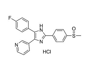
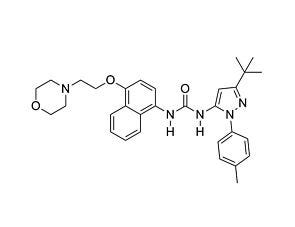
SB202190

p38 MAPK inhibitor

SB202190

p38 MAPK inhibitor
From: 238 USD
Catalog #
(Select a product)
p38 MAPK inhibitor
Add to Wish List

Overview

SB202190 is a selective, potent, cell-permeable inhibitor of p38 MAP kinases, inhibiting p38α (SAPK2A, MAPK14) and p38β (SAPK2B, MAPK11) with IC₅₀ values of 50 and 100 nM, respectively (Davies et al.; Jiang et al.). As a pyridinyl imidazole inhibitor, SB202190 directly binds p38 MAP kinases in the ATP binding pocket (Fox et al.).

MAINTENANCE AND SELF-RENEWAL
· Improves the self-renewal ability of neural stem cells from NPC1-deficient mice (Yang et al.).
· Blocks adiponectin-mediated proliferation of hematopoietic stem cells (DiMascio et al.).
· Reduces BMP3-mediated proliferation of C3H10T1/2 mesenchymal stem cells (Stewart et al.).

DIFFERENTIATION
· Induces cardiomyocyte differentiation from human embryonic stem cells (Graichen et al.).
Cell Type
Cardiomyocytes, PSC-Derived, Hematopoietic Stem and Progenitor Cells, Mesenchymal Stem and Progenitor Cells, Neural Stem and Progenitor Cells
Species
Human, Mouse, Non-Human Primate, Other, Rat
Application
Differentiation, Expansion, Maintenance
Area of Interest
Neuroscience, Stem Cell Biology
CAS Number
152121-30-7
Chemical Formula
C₂₀H₁₄FN₃O
Purity
≥ 98%
Pathway
p38 MAPK
Target
p38 MAPK

Protocols and Documentation

Find supporting information and directions for use in the Product Information Sheet or explore additional protocols below.

Document Type
Product Name
Catalog #
Lot #
Language
Product Name
SB202190
Catalog #
72634, 72632
Lot #
Lot# 1000038745 or higher for 72632 | Lot# 1000028163 or higher for 72634
Language
English
Document Type
Safety Data Sheet
Product Name
SB202190
Catalog #
72634, 72632
Lot #
All
Language
English

Applications

This product is designed for use in the following research area(s) as part of the highlighted workflow stage(s). Explore these workflows to learn more about the other products we offer to support each research area.

Resources and Publications

Publications (10)

NFATc1 deficiency in B cells ameliorates atopic dermatitis H. Kader et al. Scientific Reports 2025 Jul

Abstract

Atopic dermatitis (AD) is a common skin allergy, affecting large population worldwide. Currently, there is no cure for AD. NFATc1, a transcription factor, operates through a calcium-dependent calcineurin/calmodulin pathway to regulate target genes and is vital in immune system development and function. Previous research suggests that NFATc1 suppresses IL-10 in B cells by binding to its gene. Our current study explores the role of B cells deficient of NFATc1 during calcipotriol, a vitamin D analog, induced AD responses. Our data showed that Nfatc1f/f x mb1cre AD mice exhibited a diminished AD phenotype compared with WT AD mice, by reduced ear swelling, lower epidermal thickening, and fewer cellular infiltration to ear. This was evident by unaltered IgE levels. Interestingly, Nfatc1f/fxmb1cre AD mice displayed a higher percentage of IL-10-producing B220+CD5+CD1d+ Breg cells, indicating that NFATc1 deficiency promotes the differentiation of B cells into Bregs that produce more anti-inflammatory IL-10, thus alleviating AD symptoms. At the transcriptome level, NFATc1 deficient B cells bearing AD mice exhibited distinct gene expression profiles compared with WT AD mice, with genes that promoted B-cell development and enhanced stress and stimulus responses. This study highlights the potential of targeting NFATc1 as a molecular strategy to reduce AD symptoms without impairing B-cell function while boosting the production of the endogenous anti-inflammatory IL-10.Supplementary InformationThe online version contains supplementary material available at 10.1038/s41598-025-11247-9.
Wooden breast myopathy is characterized by satellite cell dysfunction and syndecan-4 shedding L. Pejšková et al. Frontiers in Physiology 2024 Dec

Abstract

IntroductionSkeletal muscle satellite cells (MuSCs or stem cells) play a crucial role in muscle development, maintenance, and regeneration, supporting both hypertrophy and regenerative myogenesis. Syndecans (SDCs) act as communication bridges within the muscle microenvironment, regulating interactions with extracellular matrix components and contributing significantly to tissue repair and inflammation. Specifically, syndecan-4 (SDC4) is involved in muscle regeneration at multiple stages.MethodsThis study delves into the emerging challenge of wooden breast (WB) myopathy and its connection with SDC4. Our hypothesis proposes that disruptions in MuSC dynamics through SDC4 contribute to the increased incidence of breast myopathies observed in growing broilers. To test our hypothesis, non-affected and affected broilers were systematically selected, and the characteristics of WB myopathy were studied both in vitro and in vivo. SDC4 overexpression in MuSCs and blocking peptides (BPs) corresponding to the SDC4 ectodomain were used for investigating the role of SDC4 in muscle development and its shedding levels.Results and discussion In vivo examination of affected muscles revealed smaller fibers and changes in metabolic pathways. In vitro studies unveiled disrupted proliferation of MuSCs in WB myopathy, accompanied by the downregulation of several muscle markers. Investigation of the potential role of SDC4 in the pathogenesis of WB myopathy revealed a decreased tendency in SDC4 gene expression and increased shedding of its ectodomain. Moreover, we showed that SDC4 overexpression is linked to reduced proliferation in MuSCs and affected myogenesis. We detected an impaired proliferation of WB-affected MuSCs, revealing critical insights into the dysfunctional state of these cells in myopathy. Additionally, by treating MuSCs with blocking peptides derived from the SDC4 ectodomain, we identified altered proliferation. Taken together, this work contributes with valuable knowledge on the molecular mechanisms underlying WB myopathy and the role of SDC4 in this chicken myopathy.
Lgr5 marks stem/progenitor cells contributing to epithelial and muscle development in the mouse esophagus L. Kostic et al. Nature Communications 2024 Aug

Abstract

The existence and function of Lgr5+ cells within the developing esophagus remains unknown. Here, we document multiple discrete Lgr5+ populations in the developing mouse esophagus, predominantly within nascent epithelial and external muscle layers. Lgr5 expression initially emerges in the developing proximal embryonic epithelium, but progressively extends distally and persists within the distal epithelium of neonates. Fate mapping and ablation analyses reveal a long-term contribution of epithelial Lgr5+ cells to esophageal organogenesis. Additionally, Lgr5-expressing cells are present in the developing external muscle layer, particularly during the development of the striated component. Fate mapping reveals a significant contribution of these embryonic Lgr5+ cells to the adult muscle layer. Embryonic Lgr5+ epithelial cells are also found to be important for regulating epithelial development, serving as a key source of Wnt6, among other ligands, to promote epithelial cell proliferation and formation of epithelial layers. These findings significantly enhance our understanding of esophageal development and shed light on the involvement of Lgr5+ stem/progenitor cells during organogenesis. Importantly, this study lays the foundation for investigating esophageal diseases related to the Lgr5+ stem/progenitor cell pool. The role of Lgr5+ cells in esophageal development has been unclear. Here, authors identify Lgr5+ cells in the developing mouse esophagus and reveal their crucial role in epithelial and muscle development.