FAQs on Human Airway Air-Liquid Interface Cultures
Find answers to frequently asked questions (FAQs) about culturing human airway epithelial cells (HAECs) at the air-liquid interface (ALI) using PneumaCult™.
General Information
Which PneumaCult™ media should I use?
The PneumaCult™ product line consists of serum- and bovine pituitary extract (BPE)-free culture media for culturing human airway epithelial cells. PneumaCult™-Ex Plus Medium is the most suitable choice for expanding primary human bronchial epithelial cells (HBECs) or human small airway epithelial cells (HSAECs) in 2D adherent culture. To differentiate HBECs or HSAECs at the air-liquid interface (ALI), use PneumaCult™-ALI Medium or PneumaCult™-ALI-S Medium, respectively. Differentiation of HBECs to three-dimensional (3D) airway organoids can be achieved using PneumaCult™ Airway Organoid Kit.
Learn more about PneumaCult™ culture media for airway epithelial cells >
What is the difference between PneumaCult™-Ex Medium and PneumaCult™-Ex Plus Medium for the culture of HAECs?
PneumaCult™-Ex Medium and PneumaCult™-Ex Plus Medium, respectively, are the first and second generation expansion media in the PneumaCult™ product line. We recommend using PneumaCult™-Ex Plus Medium as it supports more expansion at each passage (i.e. at least two more population doublings) while also maintaining robust mucociliary differentiation potential even after extended passaging (at least two additional passages), compared to other commercially available expansion media.
Find out how you can generate more airway epithelial cells for extended passages using PneumaCult™-Ex Plus Medium >
Is PneumaCult™-ALI Medium interchangeable with PneumaCult™-ALI-S Medium?
No, the media is not interchangeable. To generate the most representative cultures of the large and small airways, we recommend differentiating HBECs using PneumaCult™-ALI Medium and HSAECs using PneumaCult™- ALI-S Medium, respectively.
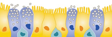
I am culturing HBECs in PneumaCult™-ALI Medium. What is the distribution of airway cell types after differentiation at air-liquid interface?
HBECs cultured in PneumaCult™-ALI Medium generate the following approximate distribution of cell types after differentiation:
- Basal cells (~20 - 30%)
- Ciliated cells (~50%)
- Goblet cells (~5 - 10%)
Studies have reported that HBECs cultured in PneumaCult™-ALI Medium can also generate rarer cell types such as ionocytes.
- Scudieri P, et al. (2020) Ionocytes and CFTR Chloride Channel Expression in Normal and Cystic Fibrosis Nasal and Bronchial Epithelial Cells. Cells 9(9):2090
I am culturing HSAECs in PneumaCult™-ALI-S Medium. What is the distribution of airway cell types after differentiation at air-liquid interface?
HSAECs cultured in PneumaCult™-ALI-S Medium generate the following approximate distribution of cell types after differentiation:
- Basal cells (~5 - 10%)
- Ciliated cells (~60 - 80%)
- Club cells (~5 - 10%)
Materials and Plate Preparation
Which Transwell® inserts should I use?
We recommend using 0.4 μm pore polyester membrane inserts to culture airway epithelial cells at the ALI using PneumaCult™-ALI Medium and PneumaCult™-ALI-S Medium. Use Costar® 6.5 mm Transwell®, 0.4 µm Pore Polyester Membrane Inserts (Catalog #38024) for 24-well plates and Costar® 12 mm Transwell®, 0.4 µm Pore Polyester Membrane Inserts (Catalog #38023) for 12-well plates—both have been validated to support optimal ALI cultures using PneumaCult™.
See data showing superior ALI culture morphology and higher epithelial cell marker expression using these recommended Transwell® inserts >
How can I scale up my ALI cultures for high-throughput applications?
Culturing HSAECs at the ALI using PneumaCult™-ALI-S Medium and HTS Transwell®-96, 0.4 µm Pore Polyester Membrane Inserts (Catalog #100-0419) can support high-throughput screening (HTS) applications up to 96-well format as reported in published literature.1 Culturing HBECs using PneumaCult™-ALI Medium and HTS Transwell®-96 performed similarly via in-house testing in 96-well plates.
Do I need to coat my inserts with collagen?
Collagen coating of Transwell® inserts and tissue culture-treated cell culture flasks is not required when expanding HAECs in PneumaCult™-Ex Plus Medium or differentiating in PneumaCult™-ALI Medium or PneumaCult™-ALI-S Medium. We have not found any performance differences between coated and non-coated conditions; however, collagen coating may improve differentiation in some donors, or if working with freshly isolated cells.
Sample Preparation
Which primary airway epithelial cell vendors have you tested for establishing ALI cultures?
There are many commercially available sources for primary airway epithelial cells. We have tested HAECs from two vendors, Lonza and Epithelix — both have resulted in successful ALI cultures.
Does PneumaCult™ support the culture of nasal epithelial cells?
Although we have not directly tested nasal epithelial cells in-house, there are publications that report PneumaCult™ supporting this cell type.
Here is a list of published literature that cites culturing of nasal epithelial cells using PneumaCult™ culture media:
Expansion using PneumaCult™-Ex Plus Medium:
- Villamizar et al. (2019) Targeted Activation of Cystic Fibrosis Transmembrane Conductance Regulator. Mol Ther 27(10): 1737–48.
Differentiation using PneumaCult™-ALI Medium:
- Müller L et al. (2013) Culturing of human nasal epithelial cells at the air liquid interface. J Vis Exp (80): 50646.
- Cao H et al. (2015) Testing gene therapy vectors in human primary nasal epithelial cultures. Mol Ther Methods Clin Dev 2: 15034.
- Schogler A et al. (2017) Characterization of pediatric cystic fibrosis airway epithelial cell cultures at the air-liquid interface obtained by non-invasive nasal cytology brush sampling. Respir Res 18(1):215.
- Garcia et al. (2019) Novel dynamics of human mucociliary differentiation revealed by single-cell RNA sequencing of nasal epithelial cultures. Development 146(20): dev177428.
- Dobzanski et al. (2018) Nasal polyp fibroblasts modulate epithelial characteristics via Wnt signaling. Int Forum Allergy Rhinol 8(12):1412–20.
Expansion and differentiation using PneumaCult™-Ex Medium and PneumaCult™-ALI Medium, respectively:
- Eckford PDW et al. (2019) The CF Canada-Sick Kids Program in individual CF therapy: A resource for the advancement of personalized medicine in CF. J Cyst Fibros 18(1):35–43.
- Cooksley C et al. (2015) TLR response pathways in NuLi-1 cells and primary human nasal epithelial cells. Mol Immunol 68(2 Pt B):476–83.
Does PneumaCult™ support the culture of murine airway epithelial cells?
Although we have not tested PneumaCult™ media for use with murine epithelial cells, there are publications that report this application.
Explore these published literature citing the culture of mourin epithelial cells using PneumaCult™-ALI Medium to learn more:
- Tata et al. (2013) Dedifferentiation of committed epithelial cells into stem cells in vivo. Nature 503(7475): 218–23.
- Johnson et al. (2018) Fank1 and Jazf1 promote multiciliated cell differentiation in the mouse airway epithelium. Biol Open 7(4): bio033944.
Setup & Culture
When should I passage my expansion cultures using PneumaCult™-Ex Plus Medium?
HBECs and HSAECs cultured in PneumaCult™-Ex Plus Medium should be passaged once they reach 50 - 60% confluence. Visualize the cellular morphology of HBEC cultures at this recommended confluence in this protocol video (skip to 02:45) >
For optimal differentiation, which passage of expanded HBECs should I use to initiate my ALI culture?
ALI cultures initiated using early passage (P3/P4) HBECs expanded in PneumaCult™-Ex Plus Medium display the most optimal morphology. Although this is donor-dependent, and some donor variability is expected, mid to late passages cultured in PneumaCult™-Ex Plus Medium can still exhibit pseudostratified mucociliary differentiation. As a precaution, we recommend not initiating a differentiation assay after P7/P8.
For optimal differentiation, which passage of expanded HSAECs should I use to initiate my ALI culture?
The best results are seen when using early passage (P3) HSAECs expanded in PneumaCult™-Ex Plus Medium. Although this is donor-dependent, and some donor variability is expected, mid-passages (P4/P5) can still have generally acceptable results. As a precaution, we recommend not initiating a differentiation assay after P5/P6.
How do I know when the submerged cultures in inserts are ready to be air-lifted?
It is critical that the submerged expansion cultures in inserts reach 100% confluence before air-lifting. At 100% confluence, the cells will cover the surface across the insert forming a complete, uniform monolayer.
View an HBEC culture that has reached the desired confluence and is ready to be air-lifted in this video (skip to 07:00) >
I seeded my cells at a higher density than that recommended in the product information sheet (PIS) and they reached confluence before 2 - 4 days. Can I proceed with air-lift?
Yes. Once your submerged culture reaches 100% confluence, it is ready to be air-lifted.
How long can I maintain my air-liquid interface culture in PneumaCult™-ALI Medium or PneumaCult™-ALI-S Medium?
Differentiated HBECs can be maintained in complete PneumaCult™-ALI Maintenance Medium for years, depending on the operators’ aseptic technique and how well they adhere to the protocol. We’ve maintained these cells for up to 5 years. HSAECs differentiated with PneumaCult™-ALI-S Medium were maintained in-house for at least 6 months.
How long does it take for HSAECs to fully differentiate?
Generally, HSAECs cultured in PneumaCult™-ALI-S Medium will form a fully differentiated cuboidal epithelium after four to five weeks of culture. Some donor variability may be expected.
When and how often should I wash the mucus off the cultures maintained in PneumaCult™-ALI Medium and PneumaCult™ALI-S Medium?
Mucus can be washed off the surface of the cells once a week starting at week 3, after the cells have been cultured in PneumaCult™-ALI Medium. Depending on the amount of mucus accumulation, a second wash may also be required. See how a mucus wash is performed in this ALI culture differentiation video (skip to 02:24) >
We find that a mucus washing step is not required for the PneumaCult™-ALI-S Medium protocol.
My culture has thicker mucus. How can I improve on the PBS wash?
To remove thicker mucus, you can add D-PBS without Ca++ and Mg++ (Catalog #37350) to the apical compartment and incubate the culture for 10 - 20 minutes at 37°C.
Cryopreservation
Can HAECs expanded in PneumaCult™-Ex Plus Medium be cryopreserved? Do you have any recommendations for freezing medium?
Yes, HAECs expanded in PneumaCult™-Ex Plus Medium can be cryopreserved. We recommend freezing them down at 1 x 10⁶ cells/mL using CryoStor® CS10 (Catalog #07930).
Characterization
How am I able to tell if the HBEC ALI differentiation assay has been successful?
There are two live-culture morphology indicators for good differentiation and readiness for further potential characterization. These are:
- visibly beating cilia at the ALI culture’s apical surface.
- visualization of secreted mucus being swept by coordinated cilia beating.
How do I assess the regional specificity of the small airway (HSAECs) vs large airway (HBECs) cultured in PneumaCult™-ALI-S Medium and PneumaCult™-ALI Medium, respectively?
To assess the regional specificity of the small vs large airway, you can perform the following assays:
- Cross-section histology followed by hematoxylin and eosin (H&E) staining to assess the thickness of the small or large airway epithelium
- Immunocytochemistry (ICC) followed by fluorescent imaging to visualize region-specific marker expression
- qPCR to evaluate differences in regional gene expression
Compare the morphology and marker expression of HSAECs and HBECs cultured in PneumaCult™-ALI-S Medium or PneumaCult™-ALI Medium in this product page (Figure 3 and 4) >
Do you have a protocol or suggested antibodies to perform ICC staining on my fully differentiated ALI cultures?
Yes, you’ll find the steps to perform an ICC staining on your epithelial cells cultured at the ALI in this protocol. Here is a list of antibodies that can be used for the characterization of airway cultures:
| Product | Catalog # | Culture Type |
|---|---|---|
| Acetylated Tubulin | e.g. 100-0753; Sigma-Aldrich, T7451 | Large and small airway ALI cultures |
| Anti-Mucin 5AC Antibody | e.g. Abcam, ab212636 | Large airway ALI cultures |
| ZO-1 Antibody | e.g. Invitrogen, 40-2200 | Large airway ALI cultures |
| CC10 Antibody (SCGB1A1) | e.g. Santa Cruz Biotechnology, sc-365992 | Small airway ALI cultures |
| Anti-SCGB3A2 Antibody | e.g. Abcam, ab181853 | Small airway ALI cultures |
Do you have a protocol for measuring transepithelial electrical resistance (TEER)? When should I perform the measurement?
Yes, you’ll find the step-by-step protocol for TEER measurement to evaluate the epithelial barrier integrity in ALI cultures here.
TEER measurements can be performed repeatedly, without causing damage to the cell culture. You can conduct a weekly TEER time course to describe the barrier function throughout the process of ALI culture differentiation. The readings can also be conducted before the culture is evaluated for endpoint characterizations, like electrophysiology or airway marker expression.
To learn how TEER values correlate with ALI culture morphology, watch this informative video.
Can't find the answer you are looking for? Reach out to us and one of our pulmonary specialists will get back to you.
Study COVID-19 with ALI Cultures
Learn how and why researchers are using ALI cultures to study respiratory viral infections, including COVID-19.

Webinar: Studying Cystic Fibrosis Using Primary Human Nasal Epithelial Cells
Explore the advantages of using nasal epithelial cell cultures, including their relevance to patients, and their implications for precision medicine in CF in this webinar. by Dr. Theo Moraes.
- Bluhmki T et al. (2020) Development of a miniaturized 96-Transwell air–liquid interface human small airway epithelial model. Sci Rep 10(1):13022
Request Pricing
Thank you for your interest in this product. Please provide us with your contact information and your local representative will contact you with a customized quote. Where appropriate, they can also assist you with a(n):
Estimated delivery time for your area
Product sample or exclusive offer
In-lab demonstration


