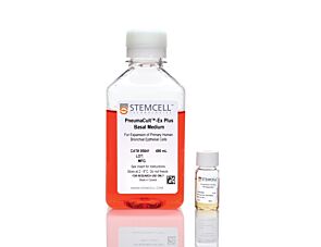
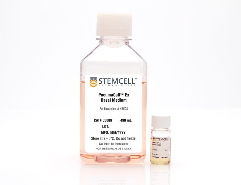
PneumaCult™-Ex Medium
Serum- and BPE-free medium for expansion of primary human airway epithelial cells
Request Pricing
Thank you for your interest in this product. Please provide us with your contact information and your local representative will contact you with a customized quote. Where appropriate, they can also assist you with a(n):
Estimated delivery time for your area
Product sample or exclusive offer
In-lab demonstration
Overview
Primary airway epithelial cells cultured in PneumaCult™-Ex expand rapidly over at least 3 passages while maintaining a cobblestone morphology and uniform expression of the basal cell markers p63 and p75NTR. Additionally, cells cultured in PneumaCult™-Ex can be differentiated to form a pseudostratified mucociliary epithelium when cultured at the air-liquid interface in PneumaCult™-ALI.
Together, PneumaCult™-Ex and PneumaCult™-ALI constitute a fully integrated BPE-free culture system for in vitro human airway modeling. This robust and defined system is a valuable tool for basic respiratory research, toxicity studies, and drug development.
Learn how to culture human airway epithelial cells at the ALI in our On-Demand Pulmonary Course or browse our Frequently Asked Questions (FAQs) about the ALI culture workflow using PneumaCult™.
Data Figures

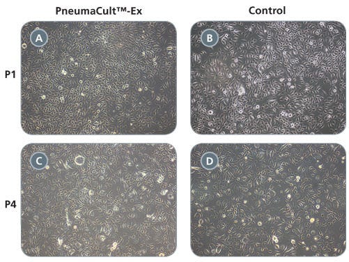
Figure 1. HBECs Cultured in PneumaCult™-Ex Exhibit Cobblestone Morphology
Commercially available, cryopreserved, passage 1 (P1) HBECs were seeded into PneumaCult™-Ex or a Control medium (BEGM™, Lonza). Cells exhibit cobblestone morphology in both culture media, as seen in representative images of confluent cultures 5 days post-seeding (A,B). HBECs cultured for an additional 3 passages in both PneumaCult™-Ex and Control medium continue to expand and retain their normal cobblestone morphology, as shown by representative images of confluent P4 cultures at 7 days post-seeding (C,D). All images were taken through 10X objective.

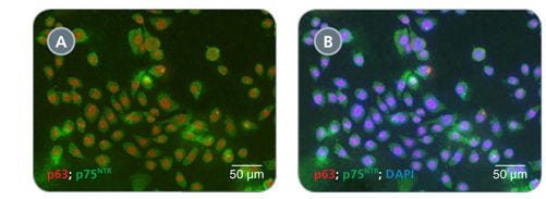
Figure 2. HBECs Cultured in PneumaCult™-Ex Exhibit Uniform Expression of Basal Cell Markers
Passage 3 HBECs cultured in PneumaCult™-Ex demonstrate extensive co-labeling of the basal cell markers p63 (red) and p75NTR (green, A). A representative merged image indicates widespread co-labeling of p63, p75NTR and the nuclear stain DAPI (blue, B).

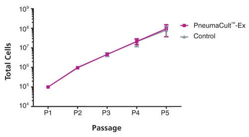
Figure 3. HBECs Cultured in PneumaCult™-Ex Exhibit Comparable Expansion Rates to Cells Cultured in Control Medium
Commercially available, cryopreserved, P1 HBECs were seeded into PneumaCult™-Ex or a Control medium (BEGM™, Lonza). In seven independent donor samples, the average fold expansion over four passages was not significantly different between cells cultured in PneumaCult™-Ex and cells cultured in the Control medium (7.1 ± 1.4 vs. 7.2 ± 1.9, mean ± SD, n = 7, p = 0.9 in paired t-test).
Protocols and Documentation
Find supporting information and directions for use in the Product Information Sheet or explore additional protocols below.
Applications
This product is designed for use in the following research area(s) as part of the highlighted workflow stage(s). Explore these workflows to learn more about the other products we offer to support each research area.
Resources and Publications
Educational Materials (10)
Publications (14)
Abstract
Abstract
Abstract
Quality Statement:
PRODUCTS ARE FOR RESEARCH USE ONLY AND NOT INTENDED FOR HUMAN OR ANIMAL DIAGNOSTIC OR THERAPEUTIC USES UNLESS OTHERWISE STATED. FOR ADDITIONAL INFORMATION ON QUALITY AT STEMCELL, REFER TO WWW.STEMCELL.COM/COMPLIANCE.