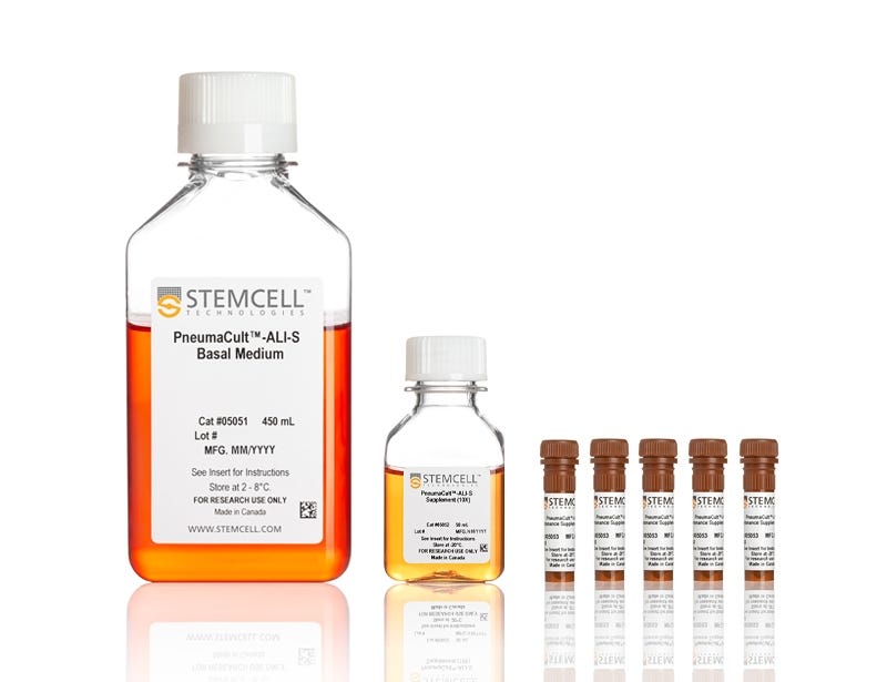
PneumaCult™-ALI-S Medium
Serum- and BPE-free medium for human small airway epithelial cells cultured at the air-liquid interface
Request Pricing
Thank you for your interest in this product. Please provide us with your contact information and your local representative will contact you with a customized quote. Where appropriate, they can also assist you with a(n):
Estimated delivery time for your area
Product sample or exclusive offer
In-lab demonstration
-
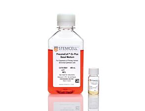 PneumaCult™-Ex Plus Medium
Serum- and BPE-free medium for expansion of primary human airway epithelial cells
-
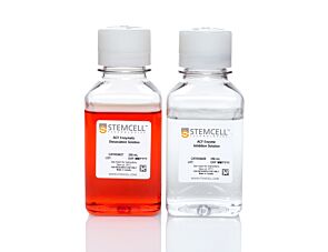 Animal Component-Free Cell Dissociation Kit
Dissociation kit for human stem and progenitor cells
-
 Hydrocortisone Stock Solution
Cell culture supplement
-
 Heparin Solution
Cell culture supplement
Overview
More Information
| Safety Statement | CA WARNING: This product can expose you to chemicals including Nickel Compounds which are known to the State of California to cause cancer and birth defects or other reproductive harm. For more information go to www.P65Warnings.ca.gov |
|---|
Data Figures
Protocols and Documentation
Find supporting information and directions for use in the Product Information Sheet or explore additional protocols below.
Applications
This product is designed for use in the following research area(s) as part of the highlighted workflow stage(s). Explore these workflows to learn more about the other products we offer to support each research area.
Resources and Publications
Educational Materials (19)


Publications (1)
Abstract
Related Products

PRODUCTS ARE FOR RESEARCH USE ONLY AND NOT INTENDED FOR HUMAN OR ANIMAL DIAGNOSTIC OR THERAPEUTIC USES UNLESS OTHERWISE STATED. FOR ADDITIONAL INFORMATION ON QUALITY AT STEMCELL, REFER TO WWW.STEMCELL.COM/COMPLIANCE.
CA WARNING: This product can expose you to chemicals including Nickel Compounds which are known to the State of California to cause cancer and birth defects or other reproductive harm. For more information go to www.P65Warnings.ca.gov
