STEMdiff™ Neural Crest Differentiation Kit

Cell culture kit for establishment of hPSC-derived neural crest cells
Need a high-quality cell source? Choose from our hiPSC healthy control lines, manufactured with mTeSR™ Plus.

STEMdiff™ Neural Crest Differentiation Kit

Cell culture kit for establishment of hPSC-derived neural crest cells

From: 452 USD
Catalog #
(Select a product)
Cell culture kit for establishment of hPSC-derived neural crest cells
Add to Wish List

Product Advantages


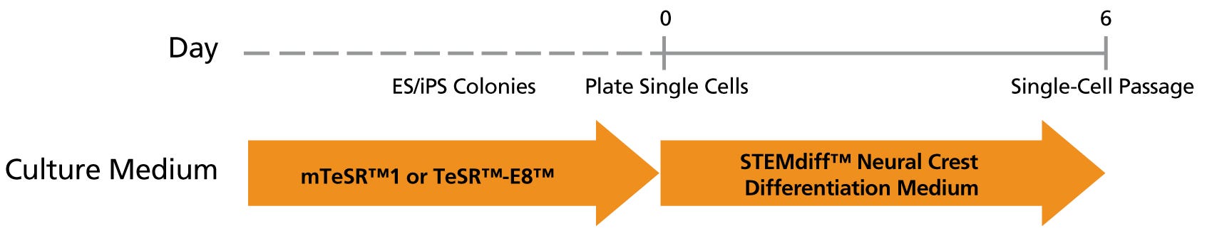
  • RAPID - Generates neural crest cells in an easy-to-use monolayer protocol in only six days

  • EFFICIENT - Greater than 70% purity of multipotent neural crest cells

  • MULTIPOTENT - Produces cells capable of downstream differentiation, including to peripheral neurons, chondrocytes, and osteoblasts

  • VERSATILE - Compatible with human ES and iPS cells maintained in mTeSR™1, mTeSR™ Plus, or TeSR™-E8™

What's Included

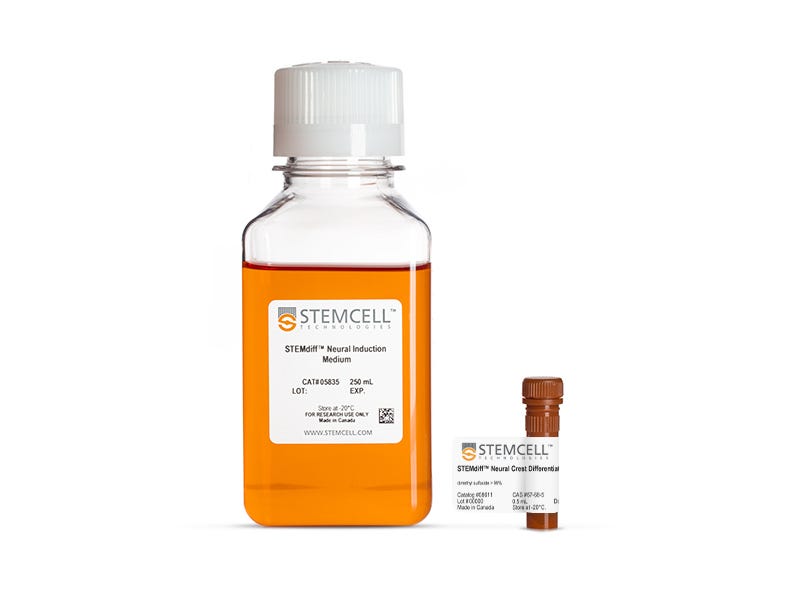
  • STEMdiff™ Neural Induction Medium, 250 mL (Catalog #05835)
  • STEMdiff™ Neural Crest Differentiation Supplement, 0.5 mL

Overview

STEMdiff™ Neural Crest Differentiation Kit creates a serum-free medium for differentiation of human embryonic stem (ES) cells and induced pluripotent stem (iPS) cells to neural crest cells. These neural crest cells, which are characterized by neural crest markers such as SOX10 and CD271, can be differentiated to several downstream derivatives including chondrocytes, osteoblasts, and peripheral neurons. This medium is compatible with human ES and iPS cells maintained in either mTeSR™1 (Catalog #85850), mTeSR™ Plus (Catalog #100-0276) or TeSR™-E8™ (Catalog #05990).
Subtype
Specialized Media
Cell Type
Neural Cells, PSC-Derived, Neural Stem and Progenitor Cells, Neurons, Pluripotent Stem Cells
Species
Human
Application
Cell Culture, Differentiation, Toxicity Assay
Brand
STEMdiff
Area of Interest
Disease Modeling, Drug Discovery and Toxicity Testing, Neuroscience, Stem Cell Biology
Formulation Category
Serum-Free

Data Figures

Figure 1.

Neural crest cells are produced using STEMdiff™ Neural Crest Differentiation Kit after 6 days in culture. For information on continued passage of neural crest cells, contact us at techsupport@stemcell.com

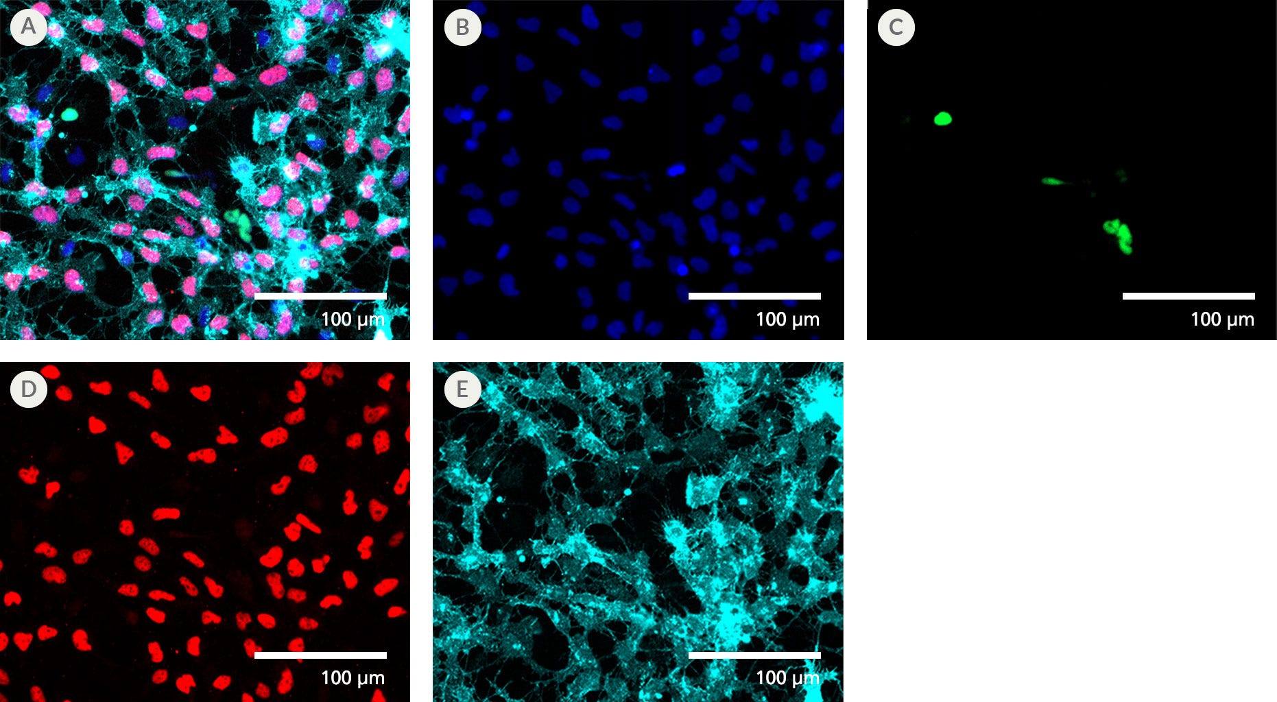
STEMdiff™ Neural Crest Differentiation Kit Generates a Highly Pure Population of NCCs with Minimal CNS-type Progenitors

Figure 2. STEMdiff™ Neural Crest Differentiation Kit Generates a Highly Pure Population of NCCs with Minimal CNS-type Progenitors

After 6 days in culture, neural crest cells (NCCs; SOX10+, red; CD271+, light blue) outnumber CNS-type progenitors (PAX6+, green). (A) Channel merge of cells fixed 2 days after being passaged on day 6. Individual immunofluorescence channels show (B) DAPI, (C) PAX6, (D) SOX10, and (E) CD271. Scale bar = 100 μm.

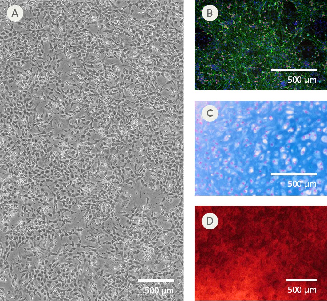
NCCs Generated with the STEMdiff™ Neural Crest Differentiation Kit Are Multipotent

Figure 3. NCCs Generated with the STEMdiff™ Neural Crest Differentiation Kit Are Multipotent

NCCs (A) were cultured for 6 days and display typical morphology. (B) Culturing NCCs using established protocols generates peripheral neurons (Peripherin, green; BRN3a, red; DAPI, blue). (C) Passaging NCCs into MesenCult™-ACF Plus Medium and then into the MesenCult™-ACF Chondrogenic Differentiation Kit generates a chondrocyte pellet (Alcian Blue, Nuclear Fast Red) with deposition of cartilage around the cells. (D) Passaging NCCs into MesenCult™-ACF Plus Medium and then into the MesenCult™ Osteogenic Differentiation Kit (Human) generates an osteoblast culture with high levels of alkaline phosphatase-positive mineral deposition. Scale bar = 500 μm (A-C), 1 mm (D).

Protocols and Documentation

Find supporting information and directions for use in the Product Information Sheet or explore additional protocols below.

Document Type
Product Name
Catalog #
Lot #
Language
Catalog #
08610
Lot #
All
Language
English
Document Type
Safety Data Sheet
Catalog #
08610
Lot #
All
Language
English

Applications

This product is designed for use in the following research area(s) as part of the highlighted workflow stage(s). Explore these workflows to learn more about the other products we offer to support each research area.

Resources and Publications

Publications (6)

Ribosomal modifications are associated with mesenchymal fate selection in the neural crest lineage I. Poverennaya et al. Nature Communications 2026 Mar

Abstract

Neural crest cells contribute to craniofacial formation by differentiating into skeletogenic mesenchyme and neuro-glial lineages. Using Smart-seq2 single-cell transcriptomics, we show that mesenchymal fate commitment correlates specifically with the expression of rRNA-modifying and ribosome assembly factors, rather than structural ribosomal proteins. Notably, EMG1 and NHP2 introduce key post-transcriptional modifications into 18S rRNA, including m¹acp³ψ at U1248, which requires TSR3 for final maturation. Disrupting NHP2 or TSR3 in vitro and in vivo perturbs cranial neural crest differentiation; post-migratory temporal knockout of Polr1a or Polr1c also causes craniofacial malformations. These findings align with cell type-specific m¹acp³ψ levels during neural crest differentiation. Given the neural crest contribution to neuroblastoma, we analyze patient data to find that elevated ribosomal control and rRNA-modifying proteins predict poorer outcomes. Complementary experiments in neuroblastoma cell lines reveal functional roles for TSR3 and WDR74 in mesenchymal-like tumor states. Together, our results link rRNA modifications and ribosome assembly to fate decisions, suggesting ribosomal heterogeneity shapes both normal development and tumor progression. Neural crest cells differentiate into skeletogenic mesenchyme and neuro-glial lineages, thereby contributing to craniofacial formation. Here, single-cell analysis of cranial neural crest shows that specific rRNA modification and ribosome assembly factors contribute to skeletogenic fate. Their disruption causes craniofacial defects, while high levels in neuroblastoma predict poor survival.
Dysregulation of sphingolipid-metabolizing enzymes in Friedreich's ataxia: In vitro and in vivo insights into therapeutic targeting. Z. Ramchunder et al. iScience 2026 Jul

Abstract

Friedreich's ataxia (FRDA) is an inherited neurodegenerative disorder caused by a GAA repeat expansion within the FXN gene, leading to reduced frataxin levels. This deficiency results in mitochondrial dysregulation, oxidative stress, and progressive cell death. Currently, only one approved treatment exists for FRDA in the United States, Canada, and the European Union, which improves neurological outcomes but has not been fully evaluated for broader disease symptoms. Therefore, identifying new therapeutic targets remains essential. Sphingolipids are increasingly recognized for their roles in neurodegeneration with emerging evidence indicating their dysregulation in FRDA. Here, we investigate whether sphingolipid-metabolizing enzymes are similarly affected and assess the therapeutic potential of targeting them. Our findings demonstrate that these enzymes are dysregulated across multiple FRDA models. Importantly, their modulation in vitro and in vivo significantly reduces mitochondrial dysfunction, enhances frataxin expression, and improves key pathological features of the disease, highlighting sphingolipid metabolism as a promising therapeutic target for FRDA.
Reaching a cell monolayer at the end of hiPSC differentiation enhances neural crest lineage commitment F. M. Duarte et al. PLOS One 2025 Sep

Abstract

Neural crest stem cells (NCSCs) compose a highly migratory, multipotent, stem cell population arising from the neural plate border of the embryonic ectoderm. Investigating the development of NCSCs is critical in understanding both embryonic development and abnormal events that underlie neurocristopathies. Suggested seeding densities in in vitro human induced pluripotent stem cells (hiPSCs) differentiation protocols, varying between 10,000 cells/cm 2 and 200,000 cells/cm 2 , demonstrate a lack of consensus on the optimal conditions to obtain NCSCs. Aiming to maximize the differentiation efficiency of hiPSCs towards the NCSCs lineage, we investigated the effect of the initial seeding density on NCSCs lineage commitment, both in fibroblast- and human peripheral blood mononuclear cell (PBMC)-derived hiPSCs. Cultures were characterized with gene and protein expression analysis assessing stemness ( OCT3/4 and NANOG ), neural crest identity ( SNAI2 and SOX10 ) and neuroectoderm identity ( PAX6 and SOX1 ). We demonstrate that reaching a confluent monolayer of cells by the end of the differentiating protocol is crucial to obtaining NCSCs from hiPSCs. To achieve this, our results indicated 17,000 cells/cm 2 is the optimal initial seeding density. Under this protocol, a confluent monolayer was reached after 8 days of differentiation and an average of 89% SOX10 positive cells were obtained. The fold change of SNAI2 and SOX10 expression was 11-fold and 17-fold higher, respectively, in cultures seeded with 17,000 cells/cm 2 , compared to the highest tested density of 200,000 cells/cm 2 . In contrast, seeding 200,000 cells/cm 2 induced neuroectoderm-like cells, confirmed by an average of 45% of cells marking positive for PAX6. With this work, we demonstrate the importance of achieving cellular confluency during NCSCs differentiation.
Need a high-quality cell source? Choose from our hiPSC healthy control lines, manufactured with mTeSR™ Plus.