Make more informed purchasing decisions with our new product availability and delivery estimate feature, now available on all product pages, in your cart, and during checkout.
Sign In
New to STEMCELL?
Register for an account to quickly and easily purchase products online and for one-click access to all educational content.
New look, same high quality and support! You may notice that your instrument or reagent packaging looks slightly different from images displayed on the website, or from previous orders. We are updating our look but rest assured, the products themselves and how you should use them have not changed. Learn more
Request Pricing
Thank you for your interest in this product.
Please provide us with your contact information and your local representative
will contact you with a customized quote. Where appropriate, they can also assist you with a(n):
Estimated delivery time for your area
Product sample or exclusive offer
In-lab demonstration
By submitting this form, you are providing your consent to STEMCELL Technologies Canada Inc. and its subsidiaries and affiliates (“STEMCELL”) to collect and use your information, and send you newsletters and emails in accordance with our privacy policy. Please contact us with any questions that you may have. You can unsubscribe or change your email preferences at any time.
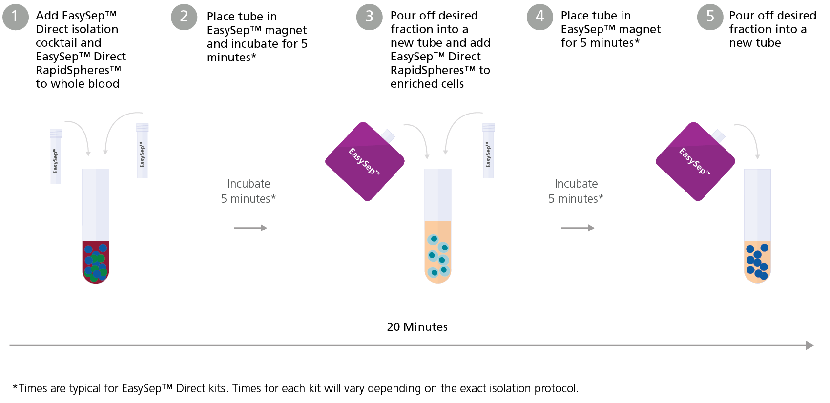
Easily and efficiently isolate highly purified human peripheral blood mononuclear cells (PBMCs) from fresh whole blood, buffy coat, bone marrow, cord blood, or leukapheresis samples by immunomagnetic negative selection, with the EasySep™ Direct Human PBMC Isolation Kit. Widely used in published research for more than 20 years, EasySep™ combines the specificity of monoclonal antibodies with the simplicity of a column-free magnetic system.
In this EasySep™ negative selection procedure, unwanted cells are labeled with antibody complexes and magnetic particles called EasySep™ Direct RapidSpheres™. The following unwanted cells are targeted for removal: granulocytes, platelets, and RBCs. The magnetically labeled cells are then separated from the untouched desired PBMCs by using an EasySep™ magnet and simply pouring or pipetting the desired cells into a new tube. Following magnetic cell isolation, the desired PBMCs are ready for downstream applications such as flow cytometry, culture, or DNA/RNA extraction.
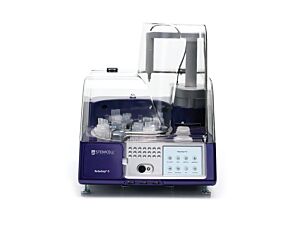
Learn more about how immunomagnetic EasySep™ technology works or how to fully automate immunomagnetic cell isolation with RoboSep™ to save time and increase laboratory throughput. Explore additional products optimized for your workflow, including those for cell characterization, cryopreservation, and more.
Magnet Compatibility
• EasySep™ Magnet (Catalog #18000)
• “The Big Easy” EasySep™ Magnet (Catalog #18001)
• EasyEights™ EasySep™ Magnet (Catalog #18103)
• Easy 50 EasySep™ Magnet (Catalog #18002)
Subtype
Cell Isolation Kits
Cell Type
B Cells, Lymphocytes, Monocytes, Mononuclear Cells, NK Cells, T Cells
Species
Human
Sample Source
Bone Marrow, Buffy Coat, Cord Blood, Leukapheresis, Whole Blood
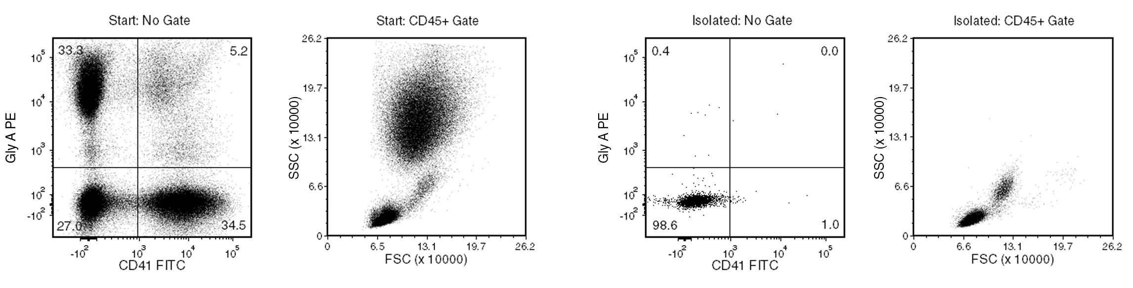
Figure 1. Typical EasySep™ Direct Human PBMC Isolation Profile
In the above example, the mononuclear cell content of the whole blood start sample (lysed by ammonium chloride) and non-lysed final isolated fraction is 27.0% and 98.6% (not gated on CD45), respectively.
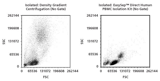
Figure 2. Representative Flow Cytometry Plots
Representative Forward Scatter (FSC) vs. Side Scatter (SSC) flow cytometry plots of mononuclear cells isolated from whole blood samples using either density gradient centrifugation or EasySep™ Direct Human PBMC Isolation Kit.
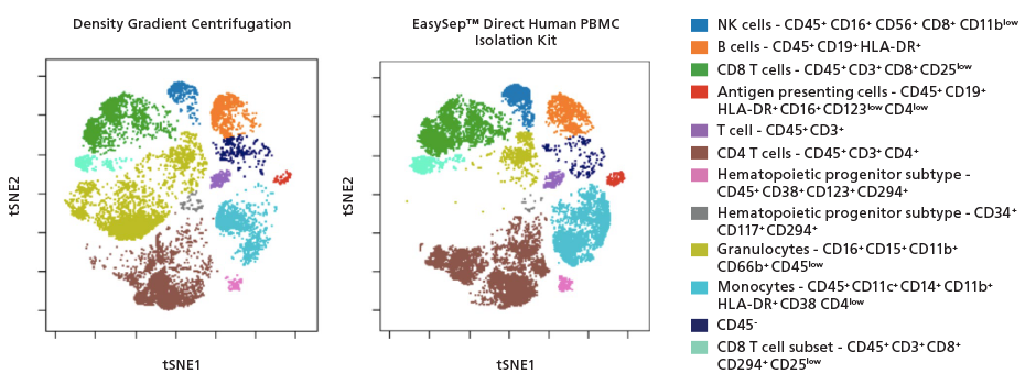
Figure 3. Representative t-SNE plots of Isolated PBMCs
Representative t-SNE plots of PBMCs stained with 19 markers and analyzed with mass cytometry (CyTOF). Cells are clustered and colored based on the combination of markers they express. Both density gradient centrifugation and EasySep™ Direct Human PBMC Isolation Kit resulted in comparable cell populations with the exception of the contaminating granulocyte population (CD16+CD15+CD11b+CD66b+ CD45low).
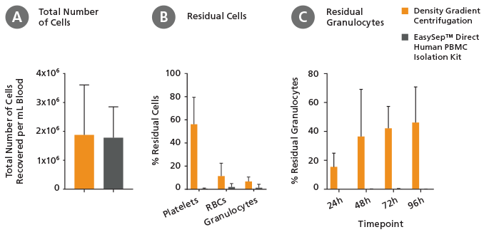
Figure 4. EasySep™ Direct Human PBMC Isolation Kit Results in Fewer Contaminating Cells Compared to Density Gradient Centrifugation
Mononuclear cells were isolated from whole blood samples using either density gradient centrifugation or EasySep™ Direct Human PBMC Isolation Kit. Cells were counted and analyzed by flow cytometry. (A) Both density gradient centrifugation and EasySep™ Direct Human PBMC Isolation Kit resulted in an equivalent total number of nucleated cells recovered from 24-hour blood samples (mean ± SD; n=14). (B) Using EasySep™ Direct Human PBMC Isolation Kit to obtain mononuclear cells from 24-hour old blood samples resulted in lower numbers of residual platelets (CD41+), red blood cells (Glycophorin A+/CD45-), and granulocytes (CD66b+) compared to density gradient centrifugation (mean ± SD; n=15). (C) Cell isolation from 24-hour, 48-hour, 72-hour and 96-hour old blood samples using EasySep™ Direct Human PBMC Isolation Kit resulted in lower numbers of residual granulocytes compared to cell isolation using density gradient centrifugation (mean ± SD; n=3).
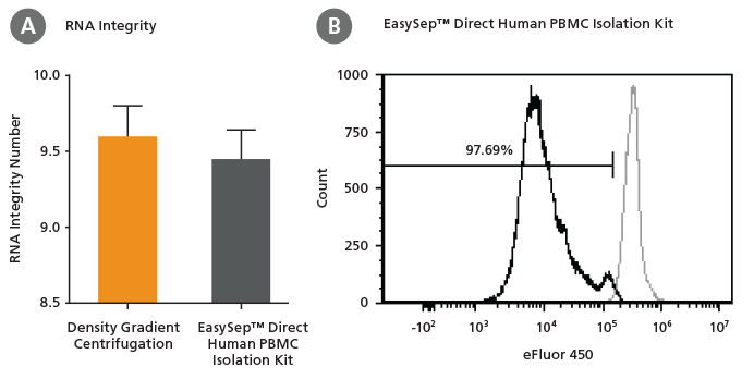
Figure 5. PBMCs Isolated with EasySep™ Direct Human PBMC Isolation Kit Proliferate and Maintain High RNA Integrity
Mononuclear cells were isolated from whole blood samples using either density gradient centrifugation or EasySep™ Direct Human PBMC Isolation Kit. (A) Isolated mononuclear cells were used for downstream RNA isolation. There was no significant difference in RNA integrity as measured with the Agilent RNA Bioanalyzer (mean ± SEM, n=3). (B) Isolated mononuclear cells were labeled with Proliferation Dye eFluor 450 and stimulated with ImmunoCult™ Human CD3/CD28 T Cell Activator and 0.5ng/ml IL-2. After 4 days in culture, cells were analyzed for proliferation by flow cytometry. Representative histogram showing dividing cells (eFluor 450low).
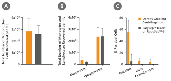
Figure 6. PBMC Isolation from a Full-Size Leukapheresis Pack Using the EasySep™ Direct Human PBMC Isolation Kit Automated with RoboSep™-S Results in Fewer Contaminating Cells Compared to Density Gradient Centrifugation
Mononuclear cells were isolated from single concentrated leukapheresis packs using either density gradient centrifugation with Lymphoprep™ density gradient medium (Density Gradient Centrifugation) or EasySep™ Direct Human PBMC Isolation Kit automated on the RoboSep™-S instrument (EasySep™ Direct on RoboSep™-S). Cells were counted and analyzed by flow cytometry. Compared to density gradient centrifugation, EasySep™ Direct Human PBMC Isolation Kit automated on the RoboSep™-S instrument resulted in (A) equivalent numbers of total mononuclear cells, (B) equivalent numbers of total monocytes and lymphocytes and (C) lower numbers of residual platelets (CD41+), red blood cells (Glycophorin A+ /CD45− ), and granulocytes (CD66b+ ) (mean ± SD n=6).
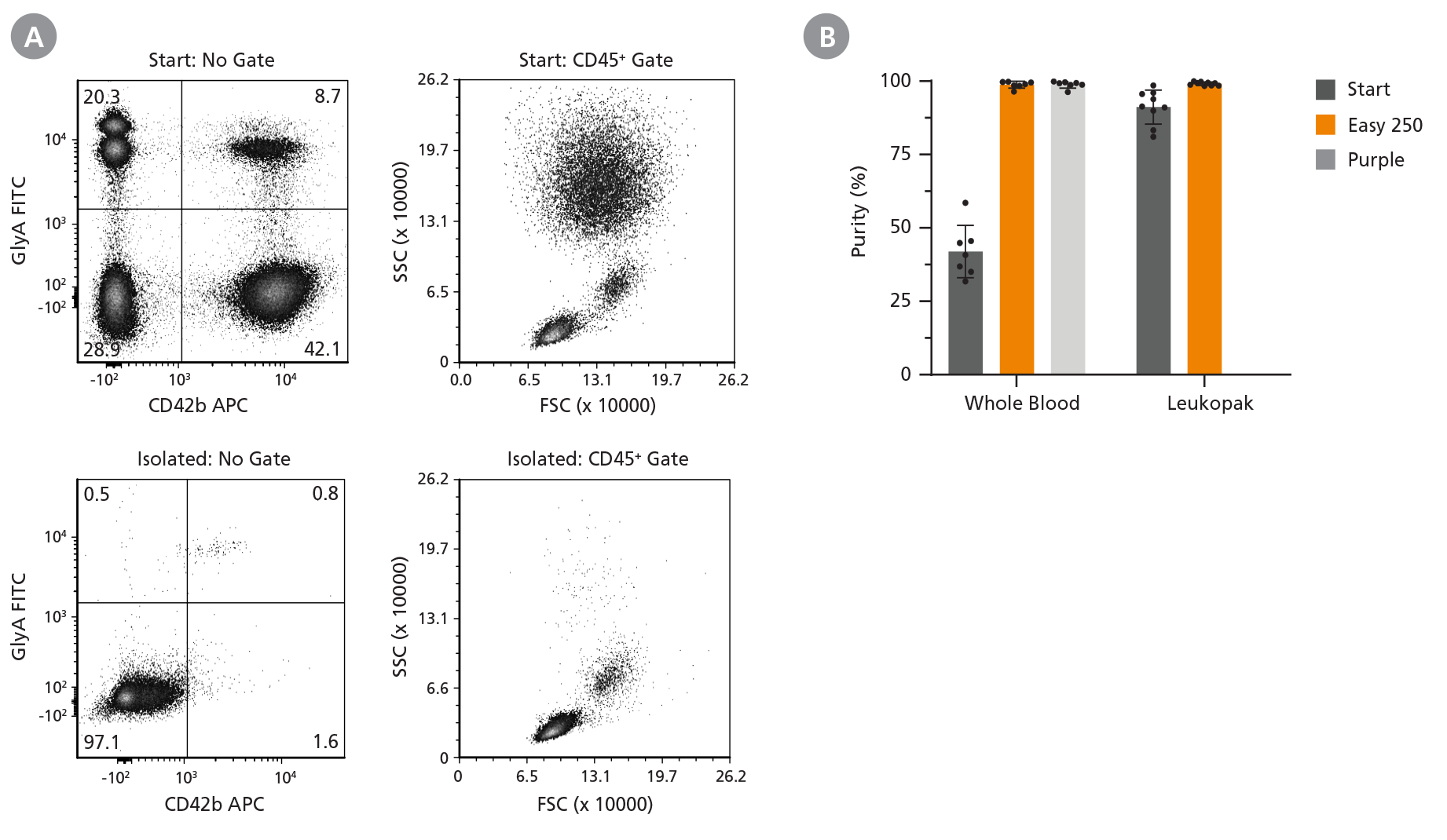
Figure 7. Easy 250 EasySep™ Magnet Can Be Used to Isolate Mononuclear Cells from Whole Blood and Leukopak Samples of Up to 125 mL
Mononuclear cells (MNCs) were isolated using the EasySep™ Direct Human PBMC Isolation Kit (Catalog #19654) from large whole blood samples (25 - 125 mL) or unprocessed leukopaks (25 - 125 mL) with the Easy 250 EasySep™ Magnet (Easy 250; Catalog #100-0821) and from small samples (1 mL whole blood) with the EasySep™ Magnet (Purple; Catalog #18000). (A) Representative flow cytometry plots show the MNC fraction (Glycophorin A- CD42b-). In the above example, the mononuclear cell content of the whole blood start sample (lysed by ammonium chloride) and non-lysed final isolated fraction is 28.9% and 97.1% (not gated on CD45), respectively. (B) Cell purity was measured before (Start) and after isolation (Easy 250, Purple) based on viable cells. Cells were counted and analyzed by flow cytometry. Data shown as mean ± SD; n = 7 - 9.
This product is designed for use in the following research area(s) as part
of the highlighted workflow stage(s). Explore these workflows to learn more about the other products we
offer to support each research area.
An additional dose of viral vector COVID-19 vaccine and mRNA COVID-19 vaccine in kidney transplant recipients: A randomized controlled trial (CVIM 4 study).
J. Bruminhent et al.
American journal of transplantation : official journal of the American Society of Transplantation and the American Society of Transplant Surgeons 2022 nov
Abstract
Immunogenicity following an additional dose of Coronavirus disease 2019 (COVID-19) vaccine was investigated in an extended primary series among kidney transplant (KT) recipients. Eighty-five KT participants were randomized to receive either an mRNA (M group; n =??43) or viral vector (V group; n =??42) vaccine. Among them, 62% were male, with a median (IQR) age of 50 (43-59) years and post-transplantation duration of 46 (26-82) months. At 2??weeks post-additional dose, there was no difference in the seroconversion rate between the M and V groups (70% vs. 65%, p =??.63). A median (IQR) of anti-RBD antibody level was not statistically different between the M group compared with the V group (51.8 [5.1-591] vs. 28.5 [2.9-119.3] BAU/ml, p =??.18). Furthermore, the percentage of participants with positive SARS-CoV-2 surrogate virus neutralization test results was not statistically different between groups (20% vs. 15%, p =??.40). S1-specific T cell and RBD-specific B cell responses were also comparable between the M and V groups (230 [41-420] vs. 268 [118-510], p =??.65 and 2 [0-10] vs. 2 [0-13] spot-forming units/106 peripheral blood mononuclear cells, p =??.60). In conclusion, compared with an additional dose of viral vector COVID-19 vaccine, a dose of mRNA COVID-19 vaccine did not elicit significantly different responses in KT recipients, regarding either humoral or cell-mediated immunity. (TCTR20211102003).
SARS-CoV-2-specific humoral and cell-mediated immune responses after immunization with inactivated COVID-19 vaccine in kidney transplant recipients (CVIM 1 study).
J. Bruminhent et al.
American journal of transplantation : official journal of the American Society of Transplantation and the American Society of Transplant Surgeons 2022 mar
Abstract
Immunogenicity following inactivated SARS-CoV-2 vaccination among solid organ transplant recipients has not been assessed. Seventy-five patients (37 kidney transplant [KT] recipients and 38 healthy controls) received two doses, at 4-week intervals, of an inactivated whole-virus SARS-CoV-2 vaccine. SARS-CoV-2-specific humoral (HMI) and cell-mediated immunity (CMI) were measured before, 4 weeks post-first dose, and 2 weeks post-second dose. The median (IQR) age of KT recipients was 50 (42-54) years and 89% were receiving calcineurin inhibitors/mycophenolate/corticosteroid regimens. The median (IQR) time since transplant was 4.5 (2-9.5) years. Among 35 KT patients, the median (IQR) of anti-RBD IgG level measured by CLIA after vaccination was not different from baseline, but was significantly lower than in controls (2.4 [1.1-3.7] vs. 1742.0 [747.7-3783.0] AU/ml, p < .01) as well as percentages of neutralizing antibody inhibition measured by surrogate viral neutralization test (0 [0-0] vs. 71.2 [56.8-92.2]%, p < .01). However, the median (IQR) of SARS-CoV-2 mixed peptides-specific T cell responses measured by ELISpot was significantly increased compared with baseline (30 [4-120] vs. 12 [0-56] T cells/106 PBMCs, p = .02) and not different from the controls. Our findings revealed weak HMI but comparable CMI responses in fully vaccinated KT recipients receiving inactivated SARS-CoV-2 vaccination compared to immunocompetent individuals (Thai Clinical Trials Registry, TCTR20210226002).
Establishment of a human iPSC line (SUTCMi001-A) derived from a healthy donor.
L. Min et al.
Stem cell research 2022 aug
Abstract
This study describes the characterization of one induced pluripotent stem cell line (iPSC) from a healthy female. It is crucial to use iPSCs derived from healthy individuals as controls in genetic disease studies. Thus, we established a human iPSC cell line derived from healthy people. The iPSC cell line was generated in our lab from the peripheral blood mononuclear cells (PBMCs) of a 28-year-old girl. The generated hiPSC line is free of episomal vectors, has a normal karyotype, expresses pluripotency markers and can differentiate into three germ layers in vivo.
Mouse monoclonal IgG2b antibody against human CD235ab (glycophorin A/B)
Item added to your cart
EasySep™ Direct Human PBMC Isolation Kit
New look, same high quality and support! You may notice that your instrument or reagent packaging looks slightly different from images displayed on the website, or from previous orders. We are updating our look but rest assured, the products themselves and how you should use them have not changed. Learn more
Quality Statement:
PRODUCTS ARE FOR RESEARCH USE ONLY AND NOT INTENDED FOR HUMAN OR ANIMAL DIAGNOSTIC OR THERAPEUTIC USES UNLESS OTHERWISE STATED. FOR ADDITIONAL INFORMATION ON QUALITY AT STEMCELL, REFER TO WWW.STEMCELL.COM/COMPLIANCE.