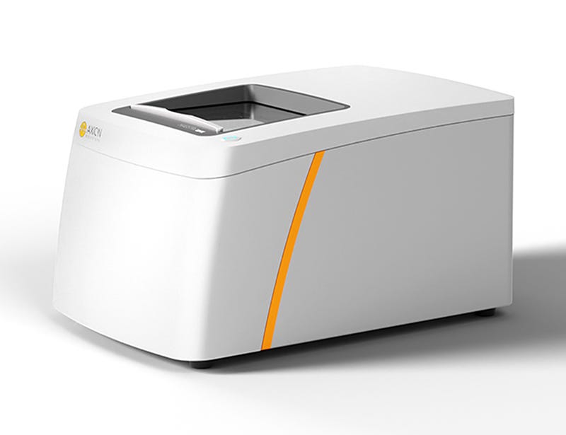
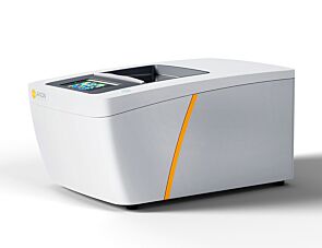
Maestro Edge™
Multiwell multi-electrode array (MEA) system with 384 channels
Request Pricing
Thank you for your interest in this product. Please provide us with your contact information and your local representative will contact you with a customized quote. Where appropriate, they can also assist you with a(n):
Estimated delivery time for your area
Product sample or exclusive offer
In-lab demonstration
-
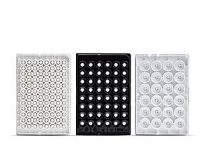 CytoView MEA™ Plate
Multiwell multi-electrode array (MEA) plate, black or white polystyrene walls with transparent SU-8 bottom; 6-, 24-, 48-, 96-well formats
-
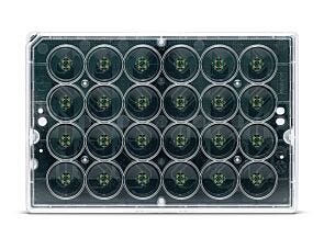 BioCircuit MEA™ Plate
Multiwell multi-electrode array (MEA) plate, clear polystyrene walls with an opaque bottom; 24-, 48-, 96-well formats
-
 Software Modules for Maestro MEA™ Systems
Data acquisition and analysis modules for Maestro Pro™ and Edge™ MEA systems
Overview
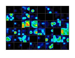
Data Figures


Protocols and Documentation
Find supporting information and directions for use in the Product Information Sheet or explore additional protocols below.
Applications
This product is designed for use in the following research area(s) as part of the highlighted workflow stage(s). Explore these workflows to learn more about the other products we offer to support each research area.
Resources and Publications
Educational Materials (15)


Related Products


PRODUCTS ARE FOR RESEARCH USE ONLY AND NOT INTENDED FOR HUMAN OR ANIMAL DIAGNOSTIC OR THERAPEUTIC USES UNLESS OTHERWISE STATED. FOR ADDITIONAL INFORMATION ON QUALITY AT STEMCELL, REFER TO WWW.STEMCELL.COM/COMPLIANCE.
