Make more informed purchasing decisions with our new product availability and delivery estimate feature, now available on all product pages, in your cart, and during checkout.
Sign In
New to STEMCELL?
Register for an account to quickly and easily purchase products online and for one-click access to all educational content.
Thank you for your interest in this product.
Please provide us with your contact information and your local representative
will contact you with a customized quote. Where appropriate, they can also assist you with a(n):
Estimated delivery time for your area
Product sample or exclusive offer
In-lab demonstration
By submitting this form, you are providing your consent to STEMCELL Technologies Canada Inc. and its subsidiaries and affiliates (“STEMCELL”) to collect and use your information, and send you newsletters and emails in accordance with our privacy policy. Please contact us with any questions that you may have. You can unsubscribe or change your email preferences at any time.
The 5E10 antibody reacts with CD90 (Thy-1), a GPI-linked membrane glycoprotein that is N-glycosylated at two sites, giving rise to 25 - 37 kDa molecules. CD90 has roles in signal transduction, cell adhesion and migration, neurite outgrowth, T cell activation, tumor suppression, and inhibition of the proliferation and differentiation of hematopoietic stem cells. It is a known ligand of β2 and β3 integrins and upregulates synthesis of fibronectin, osteonectin and thrombospondin. CD90 is broadly expressed, being found on human thymocytes, neurons, some glial cells, fibroblasts, activated endothelial cells, some leukemia cell lines and a distinct subset (<1%) of CD3+CD4+ T cells in human peripheral blood. CD90 is also expressed by small subsets of CD34+ cells in fetal liver, umbilical cord blood, bone marrow and mobilized peripheral blood cells. CD90 is considered an important marker for hematopoietic stem and progenitor cells and, in combination with other markers such as CD34, is useful to identify and isolate these cells by FACS.
This antibody clone has been verified for labeling human mesenchymal cells grown in MesenCult™ Proliferation Kit (Human; Catalog #05411) and MesenCult™-XF Medium (Catalog #05420).
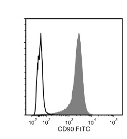
Flow cytometry analysis of human erythroleukemia (HEL) cells labeled with Anti-Human CD90 Antibody, Clone 5E10, followed by Goat Anti-Mouse IgG (H+L) Antibody, Polyclonal, FITC (Catalog #60138FI) (filled histogram), or a mouse IgG1, kappa isotype control antibody, followed by Goat Anti-Mouse IgG (H+L) Antibody, Polyclonal, FITC (solid line histogram).
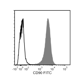
Figure 2. Data for FITC-Conjugated
Flow cytometry analysis of human erythroleukemia (HEL) cells labeled with Anti-Human CD90 Antibody, Clone 5E10, FITC (filled histogram) or Mouse IgG1, kappa Isotype Control Antibody, Clone MOPC-21, FITC (Catalog #60070FI) (solid line histogram).
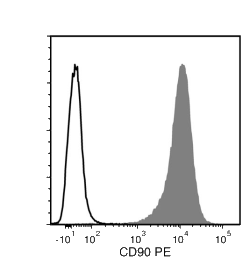
Figure 3. Data for PE-Conjugated
Flow cytometry analysis of human HEL cells labeled with Anti-Human CD90 Antibody, Clone 5E10, PE (filled histogram) or a mouse IgG1, kappa PE isotype control antibody (solid line histogram).
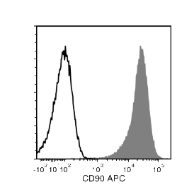
Figure 4. Data for APC-Conjugated
Flow cytometry analysis of human HEL cells labeled with Anti-Human CD90 Antibody, Clone 5E10, APC (filled histogram) or a mouse IgG1, kappa isotype control antibody, APC (solid line histogram).
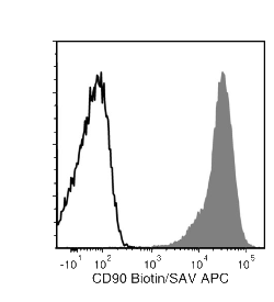
Figure 5. Data for Biotin-Conjugated
Flow cytometry analysis of human HEL cells labeled with Anti-Human CD90 Antibody, Clone 5E10, Biotin followed by streptavidin (SAV) APC (filled histogram), or a biotinylated mouse IgG1, kappa isotype control antibody followed by SAV APC (solid line histogram).
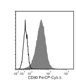
Figure 6. Data for PerCP-Cy55-Conjugated
Flow cytometry analysis of human erythroleukemina (HEL) cells labeled with Anti-Human CD90 Antibody, Clone 5E10, PerCP-Cy5.5 (filled histogram) or Mouse IgG1, kappa Isotype Control Antibody, Clone MOPC-21, PerCP-Cy5.5 (Catalog # 60070PS) (solid line histogram).
This product is designed for use in the following research area(s) as part
of the highlighted workflow stage(s). Explore these workflows to learn more about the other products we
offer to support each research area.
Intrinsic Immunity Shapes Viral Resistance of Stem Cells.
Wu X et al.
Cell 2018 JAN
Abstract
Stem cells are highly resistant to viral infection compared to their differentiated progeny; however, the mechanism is mysterious. Here, we analyzed gene expression in mammalian stem cells and cells at various stages of differentiation. We find that, conserved across species, stem cells express a subset of genes previously classified as interferon (IFN) stimulated genes (ISGs) but that expression is intrinsic, as stem cells are refractory to interferon. This intrinsic ISG expression varies in a cell-type-specific manner, and many ISGs decrease upon differentiation, at which time cells become IFN responsive, allowing induction of a broad spectrum of ISGs by IFN signaling. Importantly, we show that intrinsically expressed ISGs protect stem cells against viral infection. We demonstrate the in vivo importance of intrinsic ISG expression for protecting stem cells and their differentiation potential during viral infection. These findings have intriguing implications for understanding stem cell biology and the evolution of pathogen resistance.
Mouse monoclonal IgG1, kappa isotype control antibody
Item added to your cart
Anti-Human CD90 Antibody, Clone 5E10
Quality Statement:
PRODUCTS ARE FOR RESEARCH USE ONLY AND NOT INTENDED FOR HUMAN OR ANIMAL DIAGNOSTIC OR THERAPEUTIC USES UNLESS OTHERWISE STATED. FOR ADDITIONAL INFORMATION ON QUALITY AT STEMCELL, REFER TO WWW.STEMCELL.COM/COMPLIANCE.