TeSR™-E6

Defined, serum-free, xeno-free medium for pluripotent stem cells

TeSR™-E6

Defined, serum-free, xeno-free medium for pluripotent stem cells

From: 274 USD
Catalog #
(Select a product)
Defined, serum-free, xeno-free medium for pluripotent stem cells
Add to Wish List

What's Included

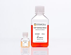
  • TeSR™-E5/E6 Basal Medium, 475 mL
  • TeSR™-E6 20X Supplement, 25 mL

Overview

TeSR™-E6 is a defined, serum- and xeno-free medium that is based on the formulation of TeSR™-E8™, but does not contain transforming growth factor β (TGF-β) or basic fibroblast growth factor (bFGF). It may be used as a basal medium for differentiation of human embryonic stem (ES) cells and induced pluripotent stem (iPS) cells, or other applications where removal of the above cytokines is desirable.
Subtype
Specialized Media
Cell Type
Pluripotent Stem Cells
Species
Human
Application
Cell Culture, Characterization, Differentiation
Brand
TeSR
Area of Interest
Stem Cell Biology
Formulation Category
Serum-Free, Xeno-Free

Protocols and Documentation

Find supporting information and directions for use in the Product Information Sheet or explore additional protocols below.

Document Type
Product Name
Catalog #
Lot #
Language
Product Name
TeSR™-E6
Catalog #
05946
Lot #
All
Language
English
Document Type
Safety Data Sheet 1
Product Name
TeSR™-E6
Catalog #
05946
Lot #
All
Language
English
Document Type
Safety Data Sheet 2
Product Name
TeSR™-E6
Catalog #
05946
Lot #
All
Language
English

Applications

This product is designed for use in the following research area(s) as part of the highlighted workflow stage(s). Explore these workflows to learn more about the other products we offer to support each research area.

Resources and Publications

Publications (12)

A human iPSC-derived 3D spinal cord organoid model to study radiation-induced neural injury. Y. Jiang et al. Stem cells translational medicine 2026 Jul

Abstract

BACKGROUND: Radiation therapy remains a cornerstone in the treatment of primary and metastatic tumors; however, its efficacy is limited by the spinal cord's high sensitivity to radiation-induced injury. To better understand the mechanisms underlying spinal cord radiosensitivity, we developed a three-dimensional human spinal cord organoid model derived from human-induced pluripotent stem cells using a neuroectodermal differentiation protocol that closely mimics embryonic spinal cord development. METHODS: Mature spinal cord organoids were exposed to a clinically relevant 2 Gy dose of ionizing radiation, and subsequent assessments included evaluation of DNA damage, astrocytic response, and neuronal functionality. RESULTS: The organoids successfully recapitulated key features of spinal cord development, including neural differentiation, spontaneous electrophysiological activity, and functional maturation. Radiation exposure led to pronounced DNA double-strand breaks, particularly in SOX2-expressing progenitor cells. Astrocyte hyperplasia was evident through increased GFAP expression, indicating a reactive gliosis response. Electrophysiological analysis revealed a marked reduction in spike frequency and burst activity, indicative of impaired neuronal function. Although partial recovery was observed over time, functional deficits persisted, suggesting sustained damage. CONCLUSION: This human spinal cord organoid model offers a physiologically relevant platform for studying radiation-induced spinal cord injury and provides valuable insights into the cellular and functional consequences of radiation exposure. It holds significant potential for advancing neuroprotective strategies and therapeutic interventions targeting radiation-induced damage in the central nervous system.
CRISPR-engineered human lung organoids with a biomolecular condensate reporter enable mechanistic toxicity monitoring S-Y. Kim et al. Materials Today Bio 2026 Feb

Abstract

Understanding how chemical stress perturbs human lung physiology requires models that capture dynamic molecular responses in real time. Here, we established a CRISPR/Cas9-engineered human induced pluripotent stem cell (hiPSC)-derived lung organoid expressing endogenous G3BP1–mCherry, enabling live, non-destructive visualization of stress granule (SG) formation under toxicant exposure. The organoids recapitulated airway and alveolar epithelial diversity and displayed lamellar body-like ultrastructures, indicating advanced maturation. Time-lapse imaging revealed rapid and reversible SG dynamics across chemically distinct stressors, while cytotoxicity assays showed that these organoids are significantly more sensitive than conventional 2D or cancer-derived lung models. Importantly, SG dynamics were linked to exposure duration–dependent changes in epithelial barrier integrity, indicating that SG formation precedes overt epithelial injury and serves as an early indicator of toxicant-induced cellular stress. Integration with high-content screening enabled quantitative, image-based analysis of cellular stress phenotypes, greatly enhancing throughput and mechanistic insight, thereby provided next-generation New Approach Methodologies for lung toxicity assessment. Together, this hiPSC-derived lung organoid SG reporter platform links early molecular stress adaptation to tissue-level responses, offering a predictive and mechanistically informative framework for human-relevant lung toxicity evaluation.
Single-cell spatial mapping of human kidney development implicates the microenvironment in guiding cell fate decisions. J. Levinsohn et al. Nature genetics 2026 Aug

Abstract

Evolution has used cell-cell communication as a strategy to coordinate organ development, enabling the reproducible generation of intricate structures. Classically, these interactions have been studied one at a time in model organisms, limiting our understanding of how cellular interplay coordinates human development. We investigated human kidney development using single-cell RNA sequencing and spatial transcriptomics, analyzing over 700,000 cells. By mapping gene expression and differentiation trajectories in space, we define the spatial organization of kidney development. Our analysis revealed unrecognized plasticity, showing that cell fate established during early patterning can be later revised. This plasticity provides a potential mechanism for how cell fate is robustly established in complex patterned tissues. Additionally, through a genome-wide, spatially aware cell-cell interaction analysis, we link localized ligand signals to cell fate decisions. We also define biologically meaningful cellular neighborhoods based on aggregated extracellular cues, providing a blueprint to understand the coordination of human development at scale.