Make more informed purchasing decisions with our new product availability and delivery estimate feature, now available on all product pages, in your cart, and during checkout.
Sign In
New to STEMCELL?
Register for an account to quickly and easily purchase products online and for one-click access to all educational content.
Thank you for your interest in this product.
Please provide us with your contact information and your local representative
will contact you with a customized quote. Where appropriate, they can also assist you with a(n):
Estimated delivery time for your area
Product sample or exclusive offer
In-lab demonstration
By submitting this form, you are providing your consent to STEMCELL Technologies Canada Inc. and its subsidiaries and affiliates (“STEMCELL”) to collect and use your information, and send you newsletters and emails in accordance with our privacy policy. Please contact us with any questions that you may have. You can unsubscribe or change your email preferences at any time.
Annexin V is a member of the annexin family of proteins that bind to membrane phospholipids in the presence of calcium. This dye has high affinity for phosphatidylserine (PS) that is present in the inner leaflet of the plasma membrane. During early-stage cell apoptosis, PS is translocated from the inner to the outer leaflet of the plasma membrane, exposing it to the external environment. Annexin V, a characteristic marker for early cell apoptosis, detects the translocation of PS to the external environment.
Annexin V is used along with viability dyes such as 7-AAD (Catalog #75001) or Propidium Iodide (Catalog #75002). The process of PS translocation occurs prior to the loss of membrane integrity. Therefore, as cells progress through apoptosis and towards necrosis, the cell membrane is compromised and consequently, viability dyes pass into the cell. Thus, cells undergoing early apoptosis stain positive for Annexin V and negative for viability dyes, while apoptotic death or necrosis is characterized by positive staining for both Annexin V and the viability dye.
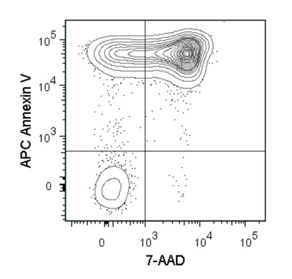
Flow cytometry analysis of C57BL/6 mouse thymocytes incubated at 37°C with 1 µM dexamethasone overnight. Cells were harvested and labeled with APC-conjugated Annexin V and 7-AAD (Catalog #75001).
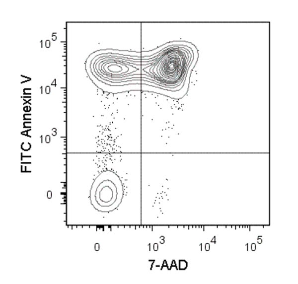
Figure 2. Data for Annexin V, FITC
Figure showing flow cytometry analysis of C57BL/6 mouse thymocytes incubated at 37°C with 1 µM dexamethasone overnight. Cells were harvested and labeled with FITC Annexin V and 7-AAD.
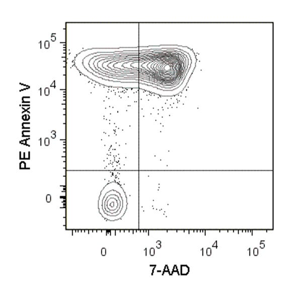
Figure 3. Data for Annexin V, PE
Figure showing flow cytometry analysis of C57BL/6 mouse thymocytes incubated at 37°C with 1 µM dexamethasone overnight. Cells were harvested and labeled with PE Annexin V and 7-AAD.
This product is designed for use in the following research area(s) as part
of the highlighted workflow stage(s). Explore these workflows to learn more about the other products we
offer to support each research area.