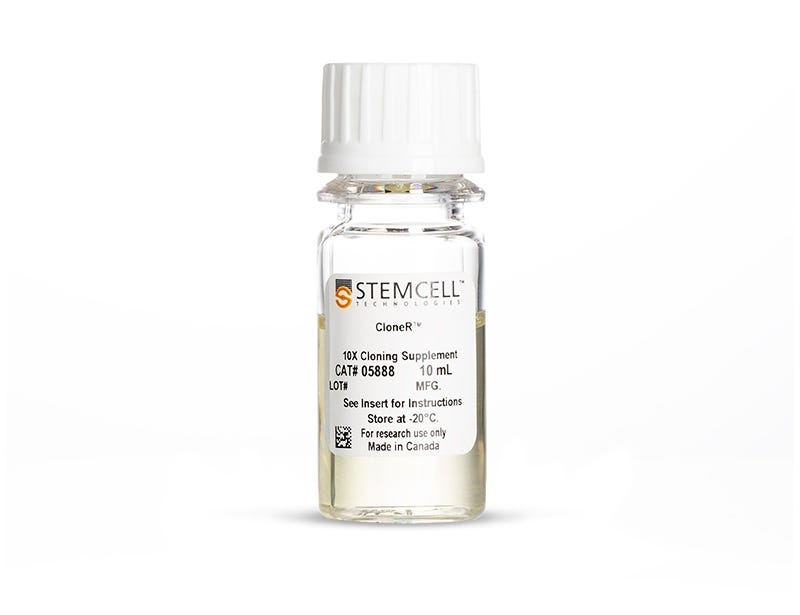
CloneR™
Defined supplement for single-cell cloning of human ES and iPS cells
Request Pricing
Thank you for your interest in this product. Please provide us with your contact information and your local representative will contact you with a customized quote. Where appropriate, they can also assist you with a(n):
Estimated delivery time for your area
Product sample or exclusive offer
In-lab demonstration
Overview
CloneR™ is compatible with the TeSR™ family of media for human ES and iPS cell maintenance as well as your choice of cell culture matrix.
For improved single-cell survival in additional applications, and higher cloning efficiency, try our CloneR™2 supplement.
Data Figures
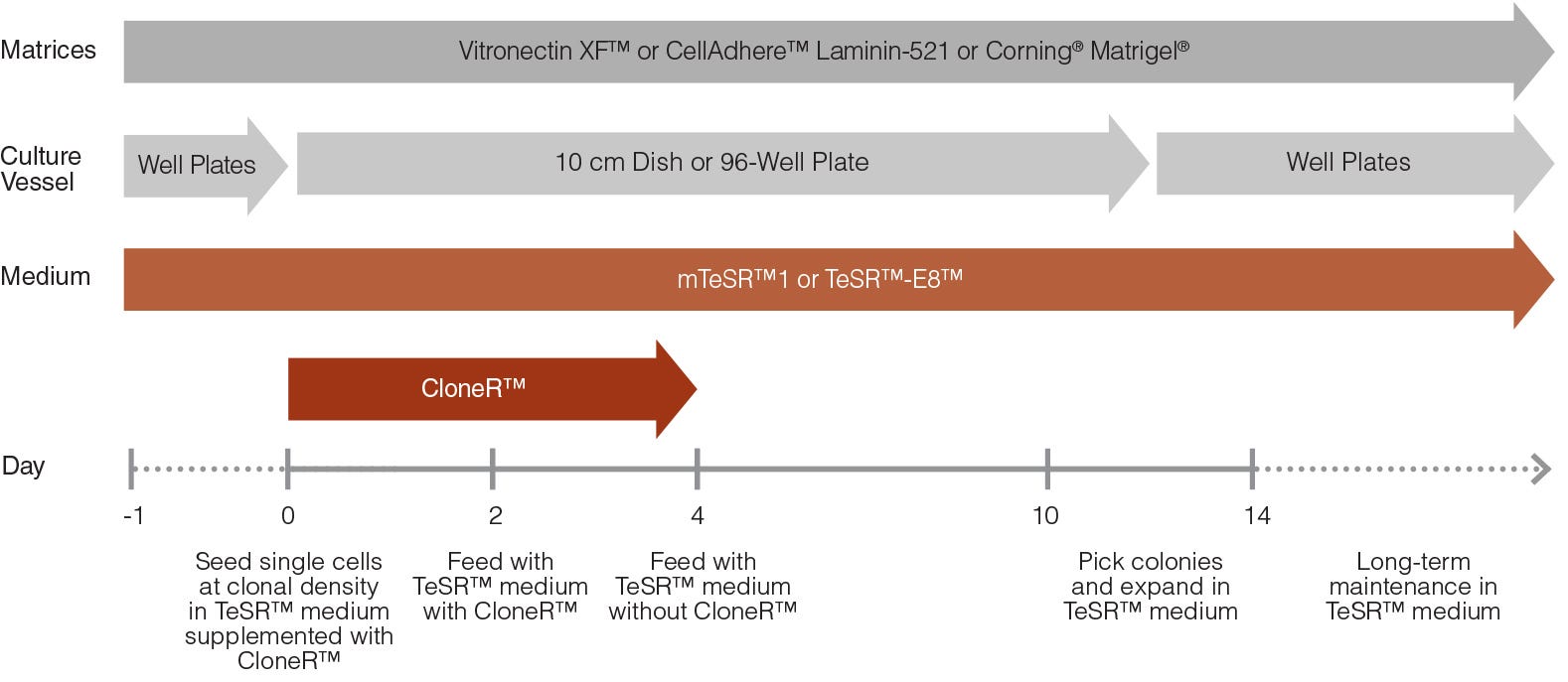
Figure 1. hPSC Single-Cell Cloning Workflow with CloneR™
On day 0, human pluripotent stem cells (hPSCs) are seeded as single cells at clonal density (e.g. 25 cells/cm2) or sorted at 1 cell per well in 96-well plates in TeSR™ (mTeSR™1 or TeSR™-E8™) medium supplemented with CloneR™. On day 2, the cells are fed with TeSR™ medium containing CloneR™ supplement. From day 4, cells are maintained in TeSR™ medium without CloneR™. Colonies are ready to be picked between days 10 - 14. Clonal cell lines can be maintained long-term in TeSR™ medium.
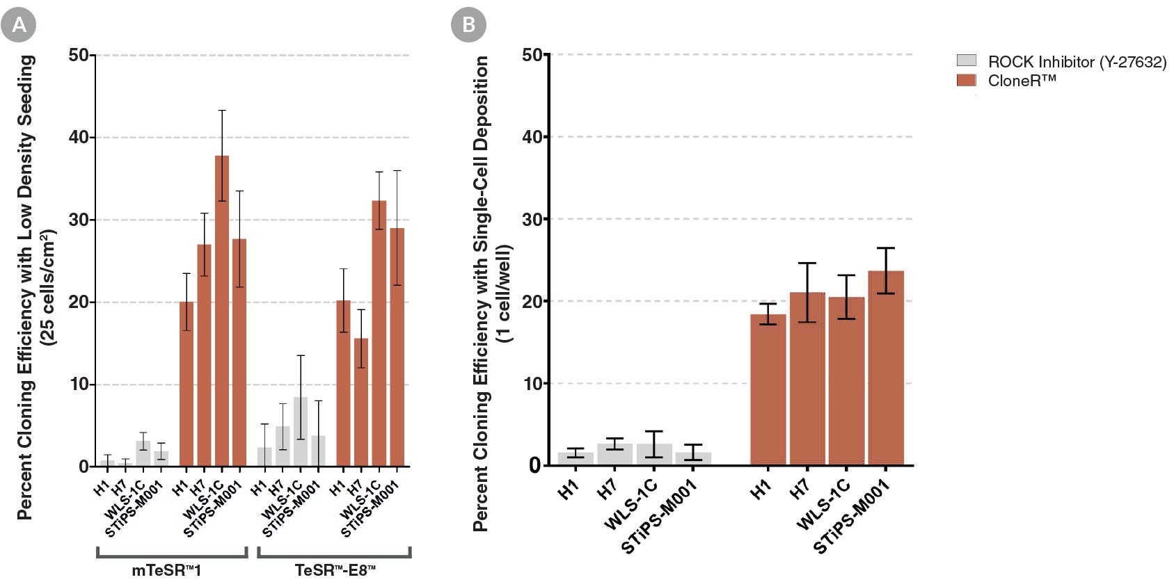
Figure 2. CloneR™ Increases the Cloning Efficiency of hPSCs and is Compatible with Multiple hPSC Lines and Seeding Protocols
TeSR™ medium supplemented with CloneR™ increases hPSC cloning efficiency compared with cells plated in TeSR™ containing ROCK inhibitor. Cells were seeded (A) at clonal density (25 cells/cm2) in mTeSR™1 and TeSR™-E8™ and (B) by single-cell deposition using FACS (seeded at 1 cell/well) in mTeSR™1.
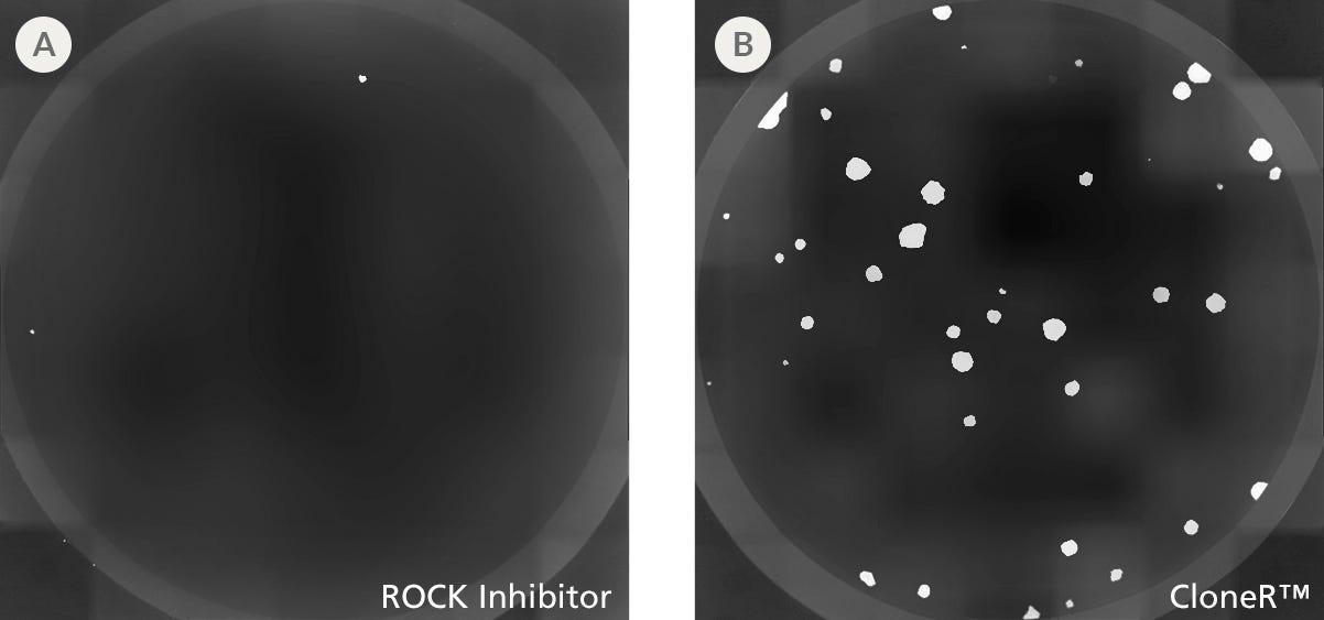
Figure 3. CloneR™ Increases the Cloning Efficiency of hPSCs at Low Seeding Densities
hPSCs plated in mTeSR™1 supplemented with CloneR™ demonstrated significantly increased cloning efficiencies compared to cells plated in mTeSR™1 containing ROCK inhibitor (10μM Y-27632). Shown are representative images of alkaline phosphatase-stained colonies at day 7 in individual wells of a 12-well plate. H1 human embryonic stem (hES) cells were seeded at clonal density (100 cells/well, 25 cells/cm2) in mTeSR™1 supplemented with (A) ROCK inhibitor or (B) CloneR™ on Vitronectin XF™ cell culture matrix.

Figure 4. CloneR™ Yields Larger Single-Cell Derived Colonies
hPSCs seeded in mTeSR™1 supplemented with CloneR™ result in larger colonies than cells seeded in mTeSR™1 containing ROCK inhibitor (10μM Y-27632). Shown are representative images of hPSC clones established after 7 days of culture in mTeSR™1 supplemented with (A) ROCK inhibitor or (B) CloneR™.
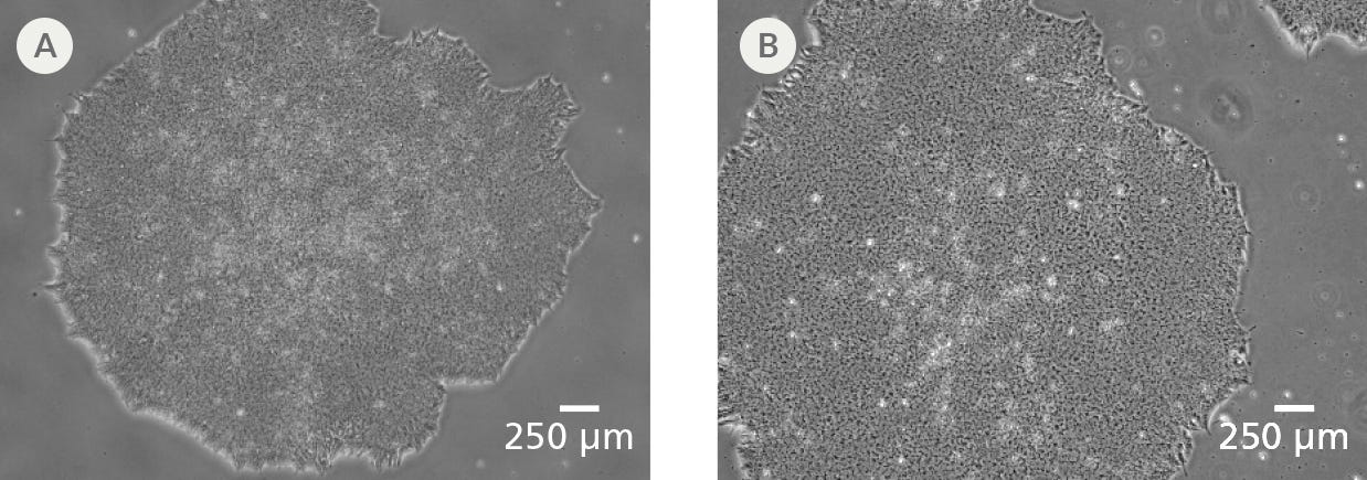
Figure 5. Clonal Cell Lines Established Using CloneR™ Display Characteristic hPSC Morphology
Clonal cell lines established using mTeSR™1 or TeSR™-E8™ medium supplemented with CloneR™ retain the prominent nucleoli and high nuclear-to-cytoplasmic ratio characteristic of hPSCs. Representative images at passage 7 after cloning are shown for clones derived from the parental (A) H1 hES cell and (B) WLS-1C human induced pluripotent stem (iPS) cell lines.
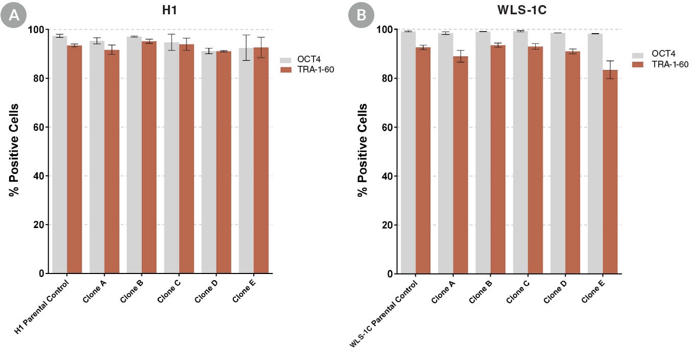
Figure 6. Clonal Cell Lines Established with CloneR™ Express High Levels of Undifferentiated Cell Markers
hPSC clonal cell lines established using mTeSR™1 supplemented with CloneR™ express comparable levels of undifferentiated cell markers, OCT4 (Catalog #60093) and TRA-1-60 (Catalog #60064), as the parental cell lines. (A) Clonal cell lines established from parental H1 hES cell line. (B) Clonal cell lines established from parental WLS-1C hiPS cell line. Data is presented between passages 5 - 7 after cloning and is shown as mean ± SEM; n = 2.
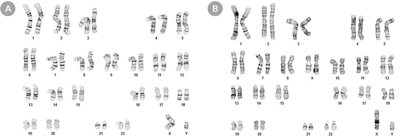
Figure 7. Clonal Cell Lines Established Using CloneR™ Display a Normal Karyotype
Representative karyograms of clones derived from parental (A) H1 hES cell and (B) WLS-1C hiPS cell lines demonstrate that the clonal lines established with CloneR™ have a normal karyotype. Cells were karyotyped 5 passages after cloning, with an overall passage number of 45 and 39, respectively.
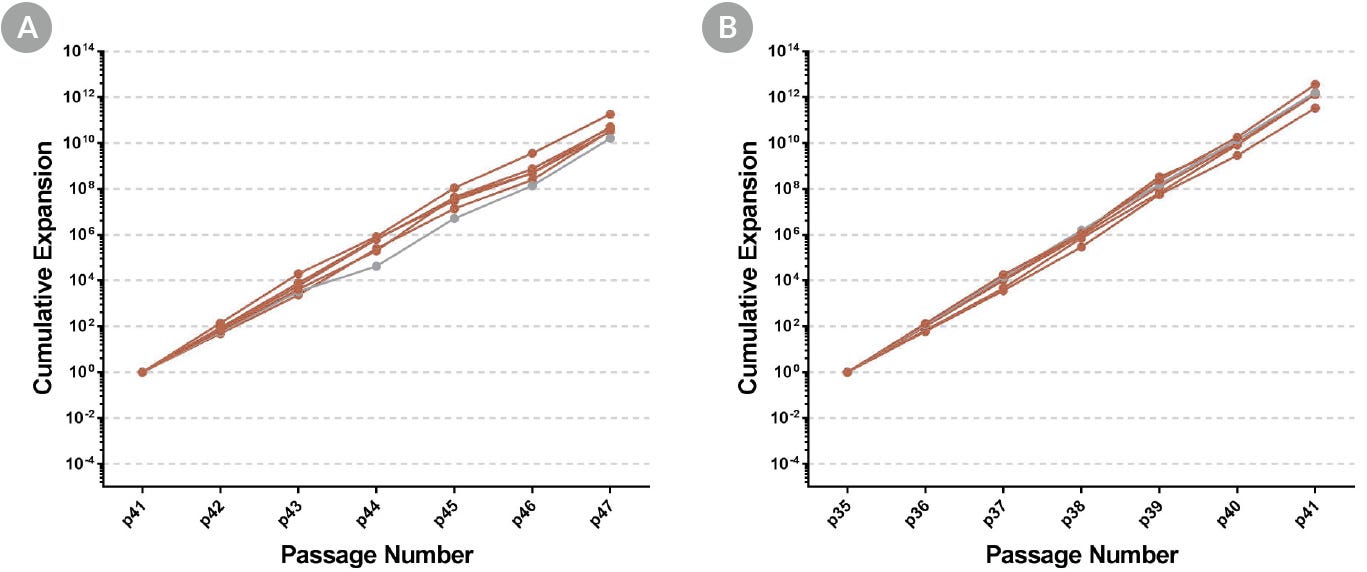
Figure 8. Clonal Cell Lines Established Using CloneR™ Display Normal Growth Rates
Fold expansion of clonal cell lines display similar growth rates to parental cell lines. Shown are clones (red) and parental cell lines (gray) for (A) H1 hES cell and (B) WLS-1C hiPS cell lines.
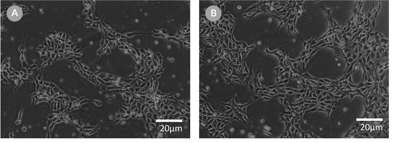
Figure 9. Representative Cell Morphology 24 Hours After RNP Electroporation in mTeSR™1 and mTeSR™ Plus
H1-eGFP ES cells were plated in (A) mTeSR™1 and (B) mTeSR™ Plus and supplemented with CloneR™ immediately following RNP electroporation. Images were taken 24 hours after electroporation.
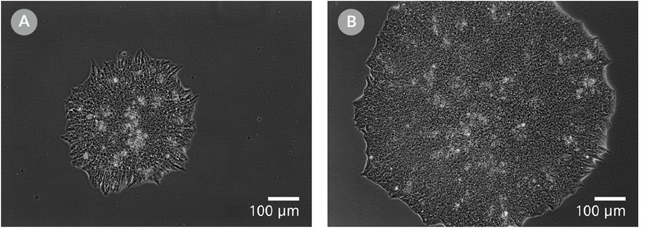
Figure 10. Clones Derived in mTeSR™ Plus are Larger and Ready to Be Picked at an Earlier Timepoint
Representative images of human ES (H9) colonies taken 8 days following singlecell plating at clonal density (25 cells/cm²) in either (A) mTeSR™1 or (B) mTeSR™ Plus supplemented with CloneR™ on CellAdhere™ Vitronectin™ XF™-coated plates.
Protocols and Documentation
Find supporting information and directions for use in the Product Information Sheet or explore additional protocols below.
Applications
This product is designed for use in the following research area(s) as part of the highlighted workflow stage(s). Explore these workflows to learn more about the other products we offer to support each research area.
Resources and Publications
Educational Materials (26)
Related Products
-
 ACCUTASE™
Cell detachment solution
-
 CloneR™2
Defined supplement for improving survival of human ES and iPS cells in single-cell workflows
-
 CellAdhere™ Laminin-521
Matrix for maintenance of human ES and iPS cells in combination with TeSR™ maintenance media
-
 mTeSR™1
cGMP, feeder-free maintenance medium for human ES and iPS cells
Item added to your cart

CloneR™
PRODUCTS ARE FOR RESEARCH USE ONLY AND NOT INTENDED FOR HUMAN OR ANIMAL DIAGNOSTIC OR THERAPEUTIC USES UNLESS OTHERWISE STATED. FOR ADDITIONAL INFORMATION ON QUALITY AT STEMCELL, REFER TO WWW.STEMCELL.COM/COMPLIANCE.
