Reversible Strainers

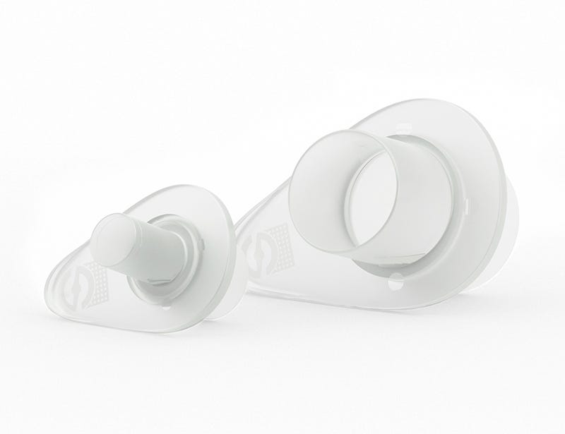
Reversible strainer for filtration of single cells and isolation of cellular aggregates, including embryoid bodies and spheroids

Reversible Strainers

Reversible strainer for filtration of single cells and isolation of cellular aggregates, including embryoid bodies and spheroids

From: 206 USD
Catalog #
(Select a product)
Reversible strainer for filtration of single cells and isolation of cellular aggregates, including embryoid bodies and spheroids
Add to Wish List

Overview

Reversible strainers are designed for filtration of single cells and isolation of cellular aggregates. They are available in 3 mesh sizes: 37 μm (white), 70 μm (orange), and 100 μm (grey). Single cells and objects smaller than the mesh will pass through the strainer and can be collected; larger aggregates will be trapped on the strainer and can be recovered separately. Strainers can be stacked for more precise size selection.

Reversible strainers can be used for harvesting embryoid bodies (EBs) and spheroids from AggreWell™ plates (e.g. Catalog #34811). Other applications include isolation of single cells, e.g. after tissue dissociation or prior to flow cytometry.

The small strainers (Catalog #27215/27216/27217) are compatible with 14 - 15 mL conical tubes (e.g. Catalog #38009) and 3 - 5 mL round-bottom tubes (e.g. Catalog #38007); the large strainers (Catalog #27250/27260/27270) are compatible with 50 mL conical tubes (e.g. Catalog #38010).
Subtype
Strainers
Species
Human, Mouse, Non-Human Primate, Other, Rat
Application
Cell Culture, Cell Isolation, Spheroid Culture

Protocols and Documentation

Find supporting information and directions for use in the Product Information Sheet or explore additional protocols below.

Document Type
Product Name
Catalog #
Lot #
Language
Catalog #
27270
Lot #
All
Language
English
Catalog #
27215
Lot #
All
Language
English
Catalog #
27260
Lot #
All
Language
English
Catalog #
27217
Lot #
All
Language
English
Catalog #
27216
Lot #
All
Language
English
Catalog #
27250
Lot #
All
Language
English

Applications

This product is designed for use in the following research area(s) as part of the highlighted workflow stage(s). Explore these workflows to learn more about the other products we offer to support each research area.

Resources and Publications

Publications (3)

A Novel Toolkit for Characterizing the Mechanical and Electrical Properties of Engineered Neural Tissues. M. Robinson et al. Biosensors 2019 apr

Abstract

We have designed and validated a set of robust and non-toxic protocols for directly evaluating the properties of engineered neural tissue. These protocols characterize the mechanical properties of engineered neural tissues and measure their electrophysical activity. The protocols obtain elastic moduli of very soft fibrin hydrogel scaffolds and voltage readings from motor neuron cultures. Neurons require soft substrates to differentiate and mature, however measuring the elastic moduli of soft substrates remains difficult to accurately measure using standard protocols such as atomic force microscopy or shear rheology. Here we validate a direct method for acquiring elastic modulus of fibrin using a modified Hertz model for thin films. In this method, spherical indenters are positioned on top of the fibrin samples, generating an indentation depth that is then correlated with elastic modulus. Neurons function by transmitting electrical signals to one another and being able to assess the development of electrical signaling serves is an important verification step when engineering neural tissues. We then validated a protocol wherein the electrical activity of motor neural cultures is measured directly by a voltage sensitive dye and a microplate reader without causing damage to the cells. These protocols provide a non-destructive method for characterizing the mechanical and electrical properties of living spinal cord tissues using novel biosensing methods.
Efficient generation of endothelial cells from human pluripotent stem cells and characterization of their functional properties Song W et al. Journal of Biomedical Materials Research - Part A 2016 OCT

Abstract

Although endothelial cells (ECs) have been derived from human pluripotent stem cells (hPSCs), large-scale generation of hPSC-ECs remains challenging and their functions are not well characterized. Here we report a simple and efficient three-stage method that allows generation of approximately 98 and 9500 ECs on day 16 and day 34, respectively, from each human embryonic stem cell (hESC) input. The functional properties of hESC-ECs derived in the presence and absence of a TGF$$-inhibitory molecule SB431542 were characterized and compared with those of human umbilical vein endothelial cells (HUVECs). Confluent monolayers formed by SB431542(+) hESC-ECs, SB431542(-) hESC-ECs, and HUVECs showed similar permeability to 10,000 Da dextran, but these cells exhibited striking differences in forming tube-like structures in 3D fibrin gels. The SB431542(+) hESC-ECs were most potent in forming tube-like structures regardless of whether VEGF and bFGF were present in the medium; less potent SB431542(-) hESC-ECs and HUVECs responded differently to VEGF and bFGF, which significantly enhanced the ability of HUVECs to form tube-like structures but had little impact on SB431542(-) hESC-ECs. This study offers an efficient approach to large-scale hPSC-EC production and suggests that the phenotypes and functions of hPSC-ECs derived under different conditions need to be thoroughly examined before their use in technology development. This article is protected by copyright. All rights reserved.
Cell Adhesion Molecules and Stem Cell-Niche-Interactions in the Limbal Stem Cell Niche. Polisetti N et al. Stem cells (Dayton, Ohio) 2016 JAN

Abstract

Interactions between stem cells and their microenvironment are critical for regulation and maintenance of stem cell function. To elucidate the molecular interactions within the human limbal epithelial stem/progenitor cell (LEPC) niche, which is essential for maintaining corneal transparency and vision, we performed a comprehensive expression analysis of cell adhesion molecules (CAMs) using custom-made quantitative real-time polymerase chain reaction (qRT-PCR) arrays and laser capture-microdissected LEPC clusters, comprising LEPCs, melanocytes, mesenchymal cells, and transmigrating immune cells. We show that LEPCs are anchored to their supporting basement membrane by the laminin receptors $\$3$\$1 and $\$6$\$4 integrin and the dystroglycan complex, while intercellular contacts between LEPCs and melanocytes are mediated by N-, P-, and E-cadherin together with L1-CAM, a member of the immunoglobulin superfamily (Ig)CAMs. In addition to the LEPC-associated heparan sulfate proteoglycans syndecan-2, glypican-3, and glypican-4, the IgCAM members ICAM-1 and VCAM-1 were found to be variably expressed on LEPCs and associated niche cells and to be dynamically regulated in response to chemokines such as interferon-$\$ enhance interactions with immune cells. Moreover, junctional adhesion molecule JAM-C accumulating in the subepithelial limbal matrix, appeared to be involved in recruitment of immune cells, while mesenchymal stromal cells appeared to use the nephronectin receptor integrin $\$8 for approaching the limbal basement membrane. In summary, we identified a novel combination of cell surface receptors that may regulate both stable and dynamic cell-matrix and cell-cell interactions within the limbal niche. The findings provide a solid foundation for further functional studies and for advancement of our current therapeutic strategies for ocular surface reconstruction.