PneumaCult™-Ex Plus Medium

Serum- and BPE-free medium for expansion of primary human airway epithelial cells
Want even better HAEC expansion rates and differentiation potential? Use PneumaCult™-NGEx Medium, our most advanced and optimized expansion medium, to achieve up to 250x more cells in just three weeks.

PneumaCult™-Ex Plus Medium

Serum- and BPE-free medium for expansion of primary human airway epithelial cells

From: 262 USD
Catalog #
(Select a product)
Serum- and BPE-free medium for expansion of primary human airway epithelial cells
Add to Wish List

Product Advantages


  • A defined, serum- and BPE-free cell culture medium that delivers consistent performance

  • PneumaCult™-Ex Plus Medium supports more cell expansion at each passage compared to other commercially available expansion media

  • When used together with PneumaCult™-ALI Medium or PneumaCult™-ALI-S Medium, PneumaCult™-Ex Plus Medium supports better ALI differentiation potential even after extended passaging compared to other commercially available expansion media

What's Included

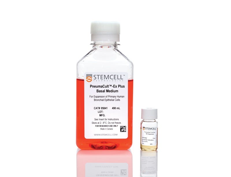
  • PneumaCult™-Ex Plus Basal Medium, 490 mL
  • PneumaCult™-Ex Plus 50X Supplement, 10 mL
Products for Your Protocol
To see all required products for your protocol, please consult the Protocols and Documentation.

Overview

PneumaCult™-Ex Plus Medium is a defined, serum- and BPE-free cell culture medium that supports more expansion of primary human airway and nasal epithelial cells at each passage, compared to other commercially available expansion media. This medium also supports at least two additional passages of cell expansion with better differentiation potential, defined as the ability to form a pseudostratified mucociliary epithelium at the air-liquid interface (ALI) using PneumaCult™-ALI Medium (Catalog #05001) or a cuboidal epithelium using PneumaCult™-ALI-S Medium (Catalog #05050).

PneumaCult™-Ex Plus and either PneumaCult™-ALI or PneumaCult™-ALI-S constitute a fully integrated BPE-free culture system for in vitro human airway modeling. This robust and defined system is a valuable tool for basic respiratory research, toxicity studies, and drug development.

Learn how to culture human airway epithelial cells at the ALI in our On-Demand Pulmonary Course or browse our Frequently Asked Questions (FAQs) about the ALI culture workflow using PneumaCult™.

For information about introductory offers to try PneumaCult™ in your lab, fill out this form.
Subtype
Specialized Media
Cell Type
Airway Cells
Species
Human
Application
Cell Culture, Expansion, Maintenance
Brand
PneumaCult
Area of Interest
Epithelial Cell Biology
Formulation Category
Serum-Free

Data Figures

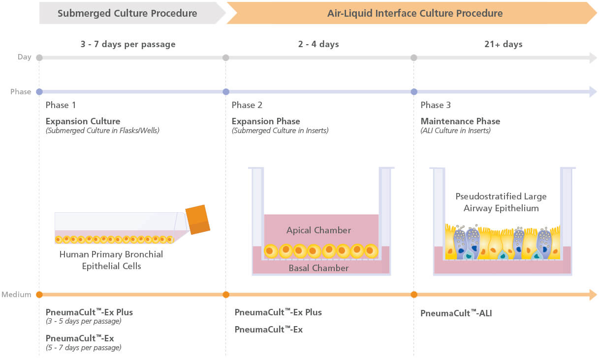
Figure 1. Overview of the PneumaCult™ culture system

Expansion of human bronchial epithelial cells (HBECs) in submerged culture is performed with PneumaCult™-Ex Plus or PneumaCult™-Ex. During the early “Expansion Phase” of the air-liquid interface (ALI) culture procedure, PneumaCult™-Ex Plus or PneumaCult™-Ex is applied to the apical and basal chambers. Upon reaching confluence, the culture is air-lifted by removing the culture medium from both chambers, and adding PneumaCult™-ALI to the basal chamber only. Differentiation into a pseudostratified mucociliary epithelium is obtained following 21-28 days of incubation and can be maintained for more than one year.

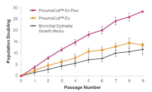
Figure 2. HBECs cultured in PneumaCult™-Ex Plus have a faster expansion rate compared to those cultured in PneumaCult™-Ex and Bronchial Epithelial Growth Media

Commercially available, cryopreserved P1 HBECs were seeded into PneumaCult™-Ex Plus, PneumaCult™-Ex, or Bronchial Epithelial Growth Media. Cells cultured in PneumaCult™-Ex Plus have a significantly higher proliferation rate over 9 passages compared to those maintained in either control medium (n=6).

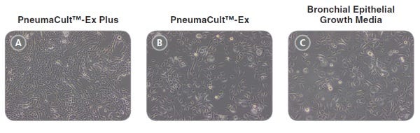
Figure 3. Representative morphology of HBECs

Representative live culture images for P4 HBECs cultured in PneumaCult™-Ex Plus, PneumaCult™-Ex, or Bronchial Epithelial Growth Media. Cells cultured in PneumaCult™-Ex Plus (A) are smaller and more tightly packed than those cultured in PneumaCult™-Ex (B) or Bronchial Epithelial Growth Media (C). All images were taken using a 10X objective.

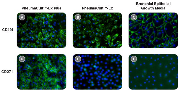
Figure 4. HBECs cultured in PneumaCult™-Ex Plus maintain widespread expression of the basal cell markers CD49f and CD271

Immunocytochemistry detection of basal cell markers - CD49f (A, B, and C) and CD271 (D, E, and F) - for P4 HBECs cultured in PneumaCult™-Ex Plus (A and D), PneumaCult™-Ex (B and E), and Bronchial Epithelial Growth Media (C and F). All images were taken using a 10X objective.

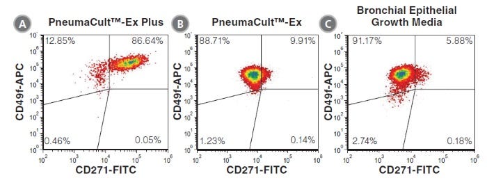
Figure 5. HBECs cultured in PneumaCult™-Ex Plus have a higher proportion of CD271+CD49f+ cells

P4 HBECs cultured in PneumaCult™-Ex Plus (A), PneumaCult™-Ex (B), and Bronchial Epithelial Growth Media (C) were characterized by flow cytometry to detect expression of the basal cell markers CD49f and CD271. HBECs cultured in PneumaCult™-Ex Plus (A) have a higher proportion of cells coexpressing CD49f and CD271, compared to those cultured in PneumaCult™-Ex (B) and Bronchial Epithelial Growth Media (C).

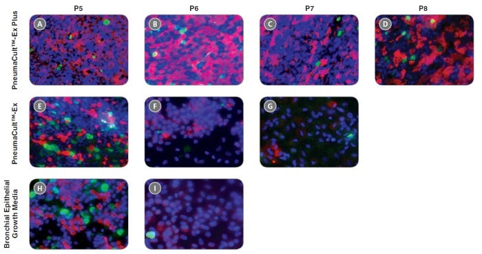
Figure 6. HBECs cultured in PneumaCult™-Ex Plus differentiate into a pseudostratified mucociliary epithelium at later passages with the use of PneumaCult™-ALI

P4 HBECs were seeded and passaged using PneumaCult™-Ex Plus, PneumaCult™-Ex, or Bronchial Epithelial Growth Media, followed by ALI differentiation at each passage (P5-8) with the use of PneumaCult™-ALI. The ALI cultures at 28 days post air-lift were fixed and stained with antibodies for cilia marker AC-tubulin (red) and the goblet cell marker Muc5AC (green). The nuclei are counterstained with DAPI (blue). All images were taken using a 20X objective.

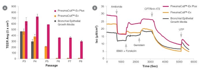
Figure 7. Electrophysiological characterization of differentiated HBECs (P4) that were expanded in PneumaCult™-Ex Plus, PneumaCult™-Ex, and Bronchial Epithelial Growth Media

Transepithelial electrical resistance (TEER) (A) and representative characterization of the ion channel activities (B) for ALI cultures at 28 days post air-lift using HBECs expanded in PneumaCult™-Ex Plus, PneumaCult™-Ex, or Bronchial Epithelial Growth Media. Amiloride: ENaC inhibitor. IBMX and Forskolin: CFTR activators. Genistein: CFTR potentiator. CFTRinh-172: CFTR inhibitor. UTP: Calciumactivated Chloride channels (CaCCs) activator. All ALI differentiation cultures were performed using PneumaCult™-ALI.

Protocols and Documentation

Find supporting information and directions for use in the Product Information Sheet or explore additional protocols below.

Document Type
Product Name
Catalog #
Lot #
Language
Catalog #
05040
Lot #
All
Language
English
Document Type
Safety Data Sheet 1
Catalog #
05040
Lot #
All
Language
English
Document Type
Safety Data Sheet 2
Catalog #
05040
Lot #
All
Language
English

Applications

This product is designed for use in the following research area(s) as part of the highlighted workflow stage(s). Explore these workflows to learn more about the other products we offer to support each research area.

Resources and Publications

Publications (61)

Blocking AGEs reduces airway epithelial viral susceptibility in diabetes-exacerbated COPD. Z. Chen et al. EMBO molecular medicine 2026 Sep

Abstract

Although diabetic complications are well-established in multiple organs, their impact on the respiratory system remains unclear. Clinical evidence indicates accelerated chronic obstructive pulmonary disease (COPD) progression in individuals with diabetes, yet the underlying mechanisms are poorly defined. Using single-cell RNA sequencing of airway epithelium, we identified distinct transcriptional alterations in COPD patients with or without diabetes, particularly in host-defense pathways related to viral infection, which is a key driver of COPD exacerbations. Functionally, hyperglycemia enhanced influenza virus susceptibility in both COPD-derived epithelial cells and COPD mice with diabetes, indicating compromised airway defense. We further demonstrated that hyperglycemia promotes advanced glycation end products (AGEs) accumulation in airway epithelium, with AGEs levels correlating with infection severity. Pharmacological inhibition of AGEs formation with aminoguanidine significantly reduced viral load both in vitro and in vivo. Mechanistically, AGEs activate p38/ERK/IKK proinflammatory signaling via RAGE and MD2 receptors, leading to epithelial injury and heightened viral susceptibility. Our findings reveal a hyperglycemia-AGEs-RAGE/MD2 axis that increases viral susceptibility in COPD, highlighting AGEs targeting as a promising therapeutic strategy for COPD patients with diabetes.
ATP/P2X7 signaling as a potential predictive biomarker for early recurrence of nasal polyps: Correlation with inflammasome activation and epithelial barrier dysfunction. L. Yang et al. The World Allergy Organization journal 2026 Sep

Abstract

BACKGROUND: Chronic rhinosinusitis with nasal polyps (CRSwNP) is characterized by pronounced epithelial remodeling and barrier dysfunction, contributing to a high rate of postoperative recurrence. However, molecular mechanisms associated with early recurrence remain poorly understood. METHODS: We performed paired proteomic profiling of nasal polyp tissues obtained from the same patients before initial surgery and at the time of recurrence (discovery cohort) to identify recurrence-associated proteins. Candidate proteins were validated in an independent cohort, and their association with recurrence risk was assessed by receiver operating characteristic (ROC) curve and Kaplan-Meier analyses. The expression and cellular localization of the identified candidate were further characterized by immunofluorescence. Mechanistic investigations were conducted in primary human nasal epithelial cells using ATP stimulation and specific pharmacological inhibitors. RESULTS: Paired proteomic analysis identified P2X7, PDE4D, and JADE2 as the top 3 upregulated proteins in recurrent tissues. Among these, only P2X7 was consistently elevated in the validation cohort and predominantly localized to the nasal epithelium (P < 0.01). ROC curve analysis indicated that tissue P2X7 level might serve as a potential predictive marker for postoperative recurrence of CRSwNP. (AUC = 0.678, P = 0.022). Kaplan-Meier analysis further showed that the risk of recurrence was significantly higher in the high P2X7 expression group (HR = 2.323, P = 0.015). Recurrent tissues exhibited elevated levels of NLRP3 and IL-1β, reduced expression of epithelial barrier markers occludin and E-cadherin, accompanied by increased extracellular ATP concentrations. In vitro, ATP stimulation upregulated P2X7 expression, induced NLRP3 and cleaved IL-1β, and downregulated occludin and E-cadherin. Notably, these effects were reversed by a P2X7-specific inhibitor (A-438079). CONCLUSION: Paired proteomic profiling revealed distinct protein expression profiles between pre- and post-recurrence nasal polyps, with P2X7 serving as a promising predictive biomarker for early recurrence. Mechanistically, the ATP/P2X7 axis may drive disease recurrence by potentially promoting inflammasome activation and epithelial barrier disruption.
Nuclear protein 1 is a cell death regulator in primary human airway epithelial cells and reduced in idiopathic pulmonary fibrosis M. Zöller et al. Scientific Reports 2026 May

Abstract

The airway epithelium is the first site of injury from cigarette smoke (CS), a major risk factor for chronic lung disease including idiopathic pulmonary fibrosis (IPF). Here, we report the first intracellular proteomic analysis of CS exposure in fully differentiated primary human bronchial epithelial cells (phBECs). Following pathway enrichment analysis, we identified nuclear protein 1 (NUPR1) as a candidate regulator of epithelial stress responses. In contrast to the prediction by pathway enrichment analysis, NUPR1 activity was not altered by CS in vitro. Nevertheless, inhibition of its nuclear translocation using ZZW-115 revealed a cytoprotective and anti-apoptotic role in phBECs, as demonstrated by increased apoptosis and impaired epithelial integrity. NUPR1 expression was markedly reduced in IPF whole lung tissue and bronchial epithelium. IPF-derived basal cells differentiated into an epithelium exhibiting fewer ciliated and more secretory cells which exhibited significantly higher sensitivity to NUPR1 inhibition. Our findings underscore cell type- and tissue-specific variation in NUPR1-dependent pathways. Collectively, this study positions NUPR1 as a context-dependent epithelial stress regulator whose loss may contribute to epithelial vulnerability in IPF.Supplementary InformationThe online version contains supplementary material available at 10.1038/s41598-026-51510-1.
Want even better HAEC expansion rates and differentiation potential? Use PneumaCult™-NGEx Medium, our most advanced and optimized expansion medium, to achieve up to 250x more cells in just three weeks.