How to Co-Culture Human Airway Epithelial and Immune Cells for RSV Infection
Respiratory syncytial virus (RSV) is a common childhood infection that can progress into severe bronchiolitis and pneumonia, making it one of the leading causes of death of infants less than 1 year of age worldwide.1 Despite this, there are relatively few therapeutic options for the treatment of RSV infection, creating a need to develop additional strategies to combat this highly prevalent disease. Recapitulating the interaction between the airway epithelium and immune cells in vitro, in the context of an RSV infection, represents an opportunity to model and advance our understanding of the mechanisms underlying disease onset and progression.
In in vitro studies, RSV can infect primary tissue-derived nasal and bronchial epithelial cells without any damage to the monolayer culture or changes in its barrier function. This suggests that the damage caused in vivo by RSV is likely a combination of viral-mediated and host-immune response pathology. In the lung, there are two sources of macrophages that may play a unique role during viral infection. First, yolk sac-derived, tissue-resident alveolar macrophages sit in the alveolar space and are mainly designated as pro-resolution macrophages with an M2-like phenotype. Second, interstitial macrophages reside in the lung parenchyma and can be recruited to the lung by appropriate inflammatory stimuli following infection. These are blood monocyte-derived macrophages that follow a molecular gradient that attracts them toward the site of infection and can present a wide phenotypic spectrum and plasticity.
To determine the role of recruited macrophages in the epithelial response to RSV infection, this protocol describes a method for the co-culture of primary tissue-derived human bronchial epithelial cells (HBECs) with monocyte-derived macrophages as a non-contact, paracrine model (Figure 1). This protocol is based on the publication by Ronaghan et al. 2022, carried out in collaboration with Theo Moraes’ lab at SickKids Hospital in Toronto.

Figure 1. Co-culturing Human Airway Epithelial Cells and Immune Cells for RSV Infection
HBECs are cultured separately at the air-liquid interface (ALI) and then placed on top of submerged macrophages to initiate co-culture. The HBECs are then infected by apical addition of medium containing RSV and incubated for 2 hours, after which the inoculum is removed and the cultures returned to ALI. The macrophages and HBECs can be collected and assessed 72 hours post-infection.
Materials
- EasySep™ Human Monocyte Isolation Kit (Catalog #19359)
- EasySep™ Magnet (Catalog #18000)
- EasySep™ Buffer (Catalog #20144)
- ImmunoCult™-SF Macrophage Medium (Catalog #10961)
- PneumaCult™-Ex Plus Medium (Catalog #05040)
- PneumaCult™-ALI Medium (Catalog #05001)
- Human Bronchial Epithelial Cells (e.g. Lonza Catalog #CC-2540S)
- Animal Component-Free Cell Dissociation Kit (Catalog #05426)
Monocyte Isolation and Differentiation into M0, M1, and M2a Macrophages
- Isolate monocytes from leukapheresis samples using EasySep™ Human Monocyte Isolation Kit.
Note: If using "The Big Easy" EasySep™ Magnet, we recommended 5 x 10⁷ cells/mL in 8 mL. Adjust as needed using EasySep™ Buffer.
- Prepare ImmunoCult™-SF Macrophage Differentiation Medium by adding Human Recombinant M-CSF to ImmunoCult™-SF Macrophage Medium to a concentration of 50 ng/mL. Mix thoroughly.
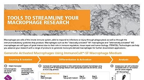
- Under "Directions for Use" in the Product Information Sheet (PIS) for ImmunoCult™-SF Macrophage Medium, follow steps 2 - 6 for a 6-day protocol using the recommended volumes and cell numbers for a 24-well plate. Differentiate into M0 macrophages, followed by M1 and M2a activation on Day 4 (Figure 2).
- Differentiate for 2 days before initiation of co-culture on Day 6.

Figure 2. Protocol for the Generation of M1 or M2a Activated Macrophages
Generate monocyte-derived macrophages (MDM) from isolated monocytes by culturing the cells in ImmunoCult™-SF Macrophage Differentiation Medium (ImmunoCult™-SF Macrophage Medium Catalog #10961 with added Human Recombinant M-CSF Catalog #78057). With our 6-day protocol, top-up with fresh ImmunoCult™-SF Macrophage Differentiation Medium and drive specific macrophage activation using appropriate stimuli (IFN-γ+LPS for M1 activation and IL-4 for M2a activation). On Day 6, initiate co-culture with fully mature M1 and M2a macrophages.
Differentiation of Human Bronchial Epithelial Cells and Co-Culture With Macrophages
- Under "Directions for Use" in the PIS for PneumaCult™-Ex Plus Medium and PneumaCult™ ALI Medium, follow the instructions provided to expand and differentiate HBECs at the air-liquid interface, following the recommended volumes and cell numbers for a 24-well plate.
- Differentiate the HBECs for 4 weeks before initiation of co-culture, and perform a medium change every 2 days.
- Prepare M0, M1, and M2a medium by adding the following concentration of cytokines to ImmunoCult™-SF Macrophage Medium:
- M0: 50 ng/mL M-CSF
- M1: 50 ng/mL M-CSF, 50 ng/mL IFNγ, 10 ng/mL LPS
- M2a: 50 ng/mL M-CSF, 10 ng/mL IL-4
- Remove media from the differentiated macrophage and HBEC cultures.
- Replace media on the basolateral side of the macrophage cultures with their respective fresh M0 medium, M1 medium, and M2a medium.
- Place Transwell® inserts containing the differentiated HBECs on top of the macrophages (Figure 1, top row).
- Incubate co-cultures for 72 hours.
- Follow the protocol outlined in How to Perform a TEER Measurement to determine the effect of co-culture on HBECs and evaluate epithelial barrier integrity in ALI cultures.
- Collect media from the cultures and evaluate for release of pro- or anti-inflammatory cytokines like TNFα, IL-10, or IL-12p70 by ELISA.
RSV Infection of Co-Cultures of HBECs and Macrophages
- Wash the apical side of the wells with 100 μL PBS.
- Replace media on the basolateral side of the co-cultures with their respective media.
- Add 0.15 MOI of RSV-GFP1 to the apical chamber and incubate at 37°C for 2 hours (Figure 1, bottom row).
Note: Multiplicity of infection (MOI) is calculated by determining the number of bronchial epithelial cells per Transwell® insert. A MOI of 1 would be one viral particle per epithelial cell. It is recommended to determine the appropriate MOI according to the donors and timeline before starting co-cultures.
- Remove the inoculum from the apical side using a pipette.
- Maintain and assess HBEC cultures for up to 72 hours post-infection (Figure 3).

Figure 3. Co-Culture of M1 Causes a Decrease in RSV Infection as Assessed by Fluorescent Signal
HBECs were placed in co-culture with M0, M1, or M2 macrophages in their respective media and infected with 0.15 MOI of RSV. Images were taken 72 hours post-infection and assessed by fluorescent signal. RSV-GFP1 is engineered to express GFP as the viral particle is being produced by the host cell. Therefore, green fluorescence is only observed when the virus is actively undergoing translation. Scale bar = 1mm.
Harvesting Macrophages for Flow Cytometry
- Collect media from the basolateral side of each well and place them in an Eppendorf tube.
- Collected media can also be used for cytokine analyses via ELISA.
- Centrifuge floating cells at 300 x g for 10 minutes. Place supernatant into a new Eppendorf tube.
Note: If needed, media can be saved for later by snap freeze.
- Add dissociation reagent to the wells as follows:
- M0 or M1 cells: 5 mL ACCUTASE™
- M2a cells: 5 mL 2.5 mM EDTA in PBS
Note: Addition of ACCUTASE™ on M2a macrophages results in loss of M2a markers2 - Incubate for 15 minutes at room temperature.
- Scrape cells with a pipette tip or mini scraper, then add to resulting pellets from floating cells in Step 2.
- Centrifuge at 300 x g for 10 minutes. Resuspend in 100 µL EasySep™ Buffer.
- Perform staining and flow cytometry with materials outlined in Table 1 to analyze viability and morphology of macrophages:
- Antibodies are added at a 1:100 dilution (see table for concentrations)
- IV3 and Rat Serum for blocking
- DRAQ7 viability stain at 1:1000 dilution
Table 1. Materials for Flow Cytometry of Macrophages
- Griffiths C et al. (2017) Respiratory syncytial virus: Infection, detection, and new options for prevention and treatment. Clin Microbiol Rev 30(1): 277–319.
- Ronaghan et al. (2022) M1-like, but not M0- or M2-like, macrophages, reduce RSV infection of primary bronchial epithelial cells in a media-dependent fashion PLoS ONE 17(10): e0276013.
- Chen et al. (2015) Impact of detachment methods on M2 macrophage phenotype and function J Immunol Methods 426:56–61.
Request Pricing
Thank you for your interest in this product. Please provide us with your contact information and your local representative will contact you with a customized quote. Where appropriate, they can also assist you with a(n):
Estimated delivery time for your area
Product sample or exclusive offer
In-lab demonstration


