Make more informed purchasing decisions with our new product availability and delivery estimate feature, now available on all product pages, in your cart, and during checkout.
Sign In
New to STEMCELL?
Register for an account to quickly and easily purchase products online and for one-click access to all educational content.
Thank you for your interest in this product.
Please provide us with your contact information and your local representative
will contact you with a customized quote. Where appropriate, they can also assist you with a(n):
Estimated delivery time for your area
Product sample or exclusive offer
In-lab demonstration
By submitting this form, you are providing your consent to STEMCELL Technologies Canada Inc. and its subsidiaries and affiliates (“STEMCELL”) to collect and use your information, and send you newsletters and emails in accordance with our privacy policy. Please contact us with any questions that you may have. You can unsubscribe or change your email preferences at any time.
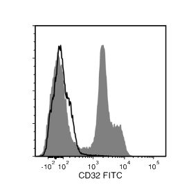
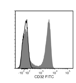
The IV.3 antibody reacts with human CD32 (FcγRII), an ~40 kDa type 1 transmembrane glycoprotein that mediates several functions including phagocytosis, cytotoxicity, immunomodulation and platelet aggregation. CD32 is encoded by three genes (A, B, C) and at least 6 isoforms are generated via alternative mRNA splicing, i.e., IIa1, IIa2, IIb1, IIb2, IIb3 and IIc. All isoforms are expressed by monocytes/macrophages, placental trophoblasts and endothelial cells. In addition, the IIb isoform is expressed by B cells, and the IIa isoform by platelets, granulocytes and, weakly, by B cells. Isoform IIc is expressed by NK cells and neutrophils. CD32 binds weakly to the Fc region of monomeric IgG but more strongly to IgG aggregates and immune complexes. These interactions can result in non-specific labeling in antibody-based detection and cell separation experiments and the IV.3 antibody may be employed as a blocking antibody to reduce non-specific binding. The IV.3 antibody binds most strongly to the IIa isoforms of CD32, with the epitope mapped to amino acids 132 - 137 [FSHLDP] in domain 2, within the ligand binding site. Binding of the IV.3 antibody can be blocked by clone FLI8.26 in flow cytometry analyses, suggesting that these clones may share a common or overlapping epitope.
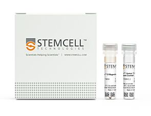
This antibody clone has been verified for purity assessments of cells isolated with EasySep™ kits, including EasySep™ Human T Cell Enrichment Kit (Catalog #19051) and EasySep™ Human CD4+ T Cell Enrichment Kit (Catalog #19052).
Subtype
Primary Antibodies
Target Antigen
CD32
Alternative Names
FCR II, FcγRII
Reactive Species
Human
Conjugation
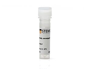
FITC, Unconjugated
Host Species
Mouse
Cell Type
B Cells, Granulocytes and Subsets, Monocytes
Species
Human
Application
Cell Isolation, Flow Cytometry, Functional Assay, Immunocytochemistry, Immunohistochemistry, Neutralization and Blocking, Western Blotting
This product is designed for use in the following research area(s) as part
of the highlighted workflow stage(s). Explore these workflows to learn more about the other products we
offer to support each research area.
Transmembrane Pickets Connect Cyto- and Pericellular Skeletons Forming Barriers to Receptor Engagement.
Freeman SA et al.
Cell 2018 JAN
Abstract
Phagocytic receptors must diffuse laterally to become activated upon clustering by multivalent targets. Receptor diffusion, however, can be obstructed by transmembrane proteins (pickets") that are immobilized by interacting with the cortical cytoskeleton. The molecular identity of these pickets and their role in phagocytosis have not been defined. We used single-molecule tracking to study the interaction between Fcγ receptors and CD44 an abundant transmembrane protein capable of indirect association with F-actin hence likely to serve as a picket. CD44 tethers reversibly to formin-induced actin filaments curtailing receptor diffusion. Such linear filaments predominate in the trailing end of polarized macrophages where receptor mobility was minimal. Conversely receptors were most mobile at the leading edge where Arp2/3-driven actin branching predominates. CD44 binds hyaluronan anchoring a pericellular coat that also limits receptor displacement and obstructs access to phagocytic targets. Force must be applied to traverse the pericellular barrier enabling receptors to engage their targets.
Mast cells form antibody-dependent degranulatory synapse for dedicated secretion and defence.
Joulia R et al.
Nature communications 2015 JAN
Abstract
Mast cells are tissue-resident immune cells that play a key role in inflammation and allergy. Here we show that interaction of mast cells with antibody-targeted cells induces the polarized exocytosis of their granules resulting in a sustained exposure of effector enzymes, such as tryptase and chymase, at the cell-cell contact site. This previously unidentified mast cell effector mechanism, which we name the antibody-dependent degranulatory synapse (ADDS), is triggered by both IgE- and IgG-targeted cells. ADDSs take place within an area of cortical actin cytoskeleton clearance in the absence of microtubule organizing centre and Golgi apparatus repositioning towards the stimulating cell. Remarkably, IgG-mediated degranulatory synapses also occur upon contact with opsonized Toxoplasma gondii tachyzoites resulting in tryptase-dependent parasite death. Our results broaden current views of mast cell degranulation by revealing that human mast cells form degranulatory synapses with antibody-targeted cells and pathogens for dedicated secretion and defence.
Mouse monoclonal IgG2b, kappa isotype control antibody
Item added to your cart
Anti-Human CD32 Antibody, Clone IV.3
Quality Statement:
PRODUCTS ARE FOR RESEARCH USE ONLY AND NOT INTENDED FOR HUMAN OR ANIMAL DIAGNOSTIC OR THERAPEUTIC USES UNLESS OTHERWISE STATED. FOR ADDITIONAL INFORMATION ON QUALITY AT STEMCELL, REFER TO WWW.STEMCELL.COM/COMPLIANCE.