Make more informed purchasing decisions with our new product availability and delivery estimate feature, now available on all product pages, in your cart, and during checkout.
Sign In
New to STEMCELL?
Register for an account to quickly and easily purchase products online and for one-click access to all educational content.
When using the EasyEights™ EasySep™ Magnet for lots 1000079626 and lower, contact us at techsupport@stemcell.com to request an additional vial of EasySep™ Dextran RapidSpheres™ 50100.
Request Pricing
Thank you for your interest in this product.
Please provide us with your contact information and your local representative
will contact you with a customized quote. Where appropriate, they can also assist you with a(n):
Estimated delivery time for your area
Product sample or exclusive offer
In-lab demonstration
By submitting this form, you are providing your consent to STEMCELL Technologies Canada Inc. and its subsidiaries and affiliates (“STEMCELL”) to collect and use your information, and send you newsletters and emails in accordance with our privacy policy. Please contact us with any questions that you may have. You can unsubscribe or change your email preferences at any time.
Isolate highly purified mouse CD45+ cells from mouse splenocytes, lung, or other tissue samples by immunomagnetic positive selection, with the EasySep™ Mouse CD45 Positive Selection Kit. Widely used in published research for more than 20 years, EasySep™ combines the specificity of monoclonal antibodies with the simplicity of a column-free magnetic system.
In this EasySep™ positive selection procedure, desired cells are labeled with antibody complexes recognizing CD45 and magnetic particles. Labeled cells are separated using an EasySep™ magnet and by simply pouring or pipetting off the unwanted cells. The cells of interest remain in the tube. Following magnetic cell isolation, the desired mouse CD45+ cells are ready for downstream applications such as flow cytometry, culture, and cell-based experiments.
Learn more about how immunomagnetic EasySep™ technology works. Explore additional products optimized for your workflow, including culture media, supplements, antibodies, and more.
Magnet Compatibility
• EasySep™ Magnet (Catalog #18000)
• “The Big Easy” EasySep™ Magnet (Catalog #18001)
• EasyEights™ EasySep™ Magnet (Catalog #18103)
• EasyPlate™ EasySep™ Magnet (Catalog #18102)
Subtype
Cell Isolation Kits
Cell Type
B Cells, Dendritic Cells, Granulocytes and Subsets, Hematopoietic Stem and Progenitor Cells, Innate Lymphoid Cells, Leukemia/Lymphoma Cells, Lymphocytes, Macrophages, Megakaryocytes, Monocytes, Mononuclear Cells, Myeloid Cells, NK Cells, Plasma, T Cells, T Cells, Other Subsets
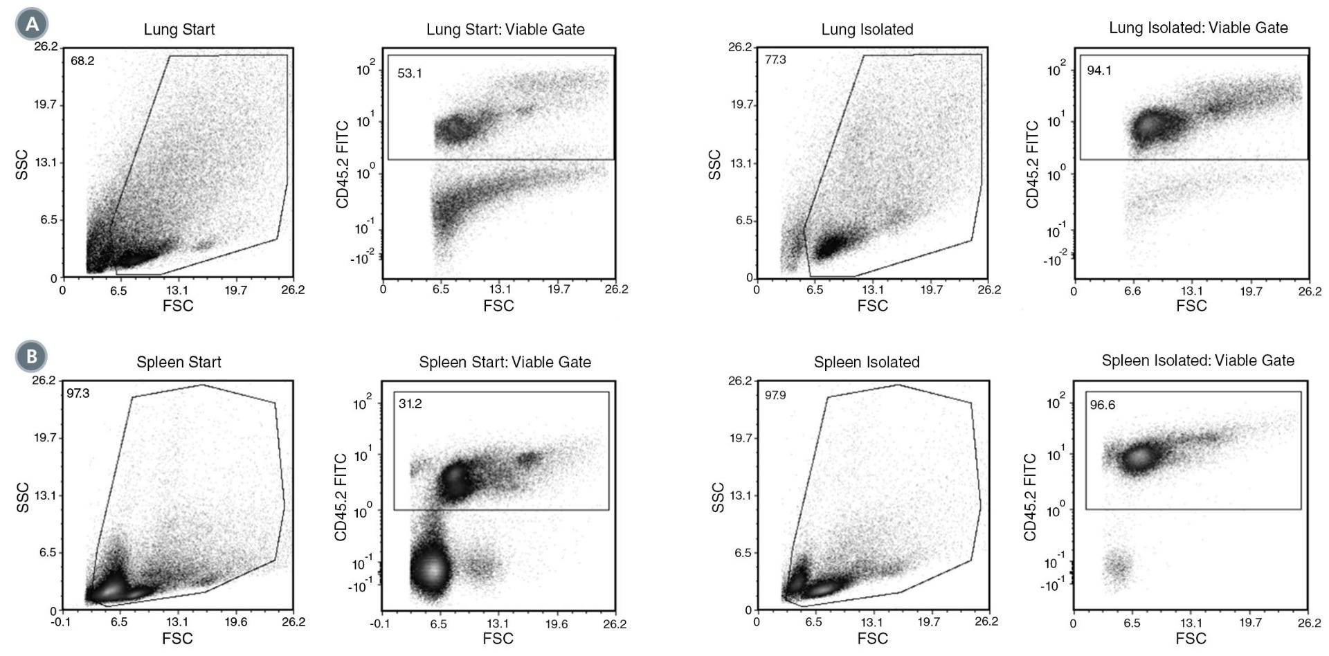
(A) Starting with a naïve mouse lung single-cell suspension, the leukocyte content (CD45+) of the isolated fraction is typically 97.0% ± 1.4% (mean ± SD) using the purple EasySep™ Magnet. In the above example, the purities of the start and final isolated fractions in lung are 53.1% and 94.1%, respectively.
(B) Starting with unlysed naïve mouse splenocytes, the leukocyte content (CD45+) of the isolated fraction is typically 97.6 ± 1.3% (mean ± SD) using the purple EasySep™ Magnet. In the above example using spleen, the purities of the start and final isolated fractions are 31.2% and 96.6%, respectively.
This product is designed for use in the following research area(s) as part
of the highlighted workflow stage(s). Explore these workflows to learn more about the other products we
offer to support each research area.
HIF-1$\alpha$ modulates sex-specific Th17/Treg responses during hepatic amoebiasis.
M. Groneberg et al.
Journal of hepatology 2022 jan
Abstract
BACKGROUND & AIMS An invasive form of intestinal Entamoeba (E.) histolytica infection, which causes amoebic liver abscess, is more common in men than in women. Immunopathological mechanisms are responsible for the more severe outcome in males. Here, we used a mouse model of hepatic amoebiasis to investigate the contribution of hepatic hypoxia-inducible factor (HIF)-1$\alpha$ to T helper 17 (Th17)/regulatory T cell (Treg) responses in the context of the sex-specific outcome of liver damage. METHODS C57BL/6J mice were infected intrahepatically with E. histolytica trophozoites. HIF-1$\alpha$ expression was determined by qPCR, flow cytometry and immunohistochemistry. Tregs and Th17 cells were analysed by immunohistochemistry and flow cytometry. Finally, male and female hepatocyte-specific Hif1$\alpha$ knockout mice were generated, and the effect of HIF-1$\alpha$ on abscess development, the cytokine milieu, and Th17/Treg differentiation was examined. RESULTS E. histolytica infection increased hepatic HIF-1$\alpha$ levels, along with the elevated frequencies of hepatic Th17 and Treg cells. While the Th17 cell population was larger in male mice, Tregs characterised by increased expression of Foxp3 in female mice. Male mice displayed increased IL-6 expression, contributing to immunopathology; this increase in IL-6 expression declined upon deletion of hepatic HIF-1$\alpha$. In both sexes, hepatic deletion of HIF-1$\alpha$ reduced the Th17 cell frequency; however, the percentage of Tregs was reduced in female mice only. CONCLUSIONS Hepatic HIF-1$\alpha$ modulates the sex-specific outcome of murine E. histolytica infection. Our results suggest that in male mice, Th17 cells can be modulated by hepatic HIF-1$\alpha$ via IL-6, indicating marked involvement in the immunopathology underlying abscess development. Strong expression of Foxp3 by hepatic Tregs from female mice suggests a potent immunosuppressive function, leading to initiation of liver regeneration. LAY SUMMARY Infection with the parasite Entamoeba histolytica activates immunopathological mechanisms in male mice, which lead to liver abscesses that are larger than those in female mice. In the absence of the protein HIF-1$\alpha$ in hepatocytes, abscess formation is reduced; moreover, the sex difference in abscess size is abolished. These results suggest that HIF-1$\alpha$ modulates the immune response involved in the induction of immunopathology, resulting in differential disease susceptibility in males and females.
Human NK cells confer protection against HIV-1 infection in humanized mice.
C. M. Sungur et al.
The Journal of clinical investigation 2022 dec
Abstract
The role of NK cells against HIV-1 infections remains to be elucidated in vivo. While humanized mouse models potentially could be used to directly evaluate human NK cell responses during HIV-1 infection, improved functional development of human NK cells in these hosts is needed. Here, we report the humanized MISTRG-6-15 mouse model, in which NK cells were quick to expand and exhibit degranulation, cytotoxicity, and proinflammatory cytokine production in nonlymphoid organs upon HIV-1 infection but had reduced functionality in lymphoid organs. Although HIV-1 infection induced functional impairment of NK cells, antiretroviral therapy reinvigorated NK cells in response to HIV-1 rebound after analytic treatment interruption. Moreover, a broadly neutralizing antibody, PGT121, enhanced NK cell function in vivo, consistent with antibody-dependent cellular cytotoxicity. Monoclonal antibody depletion of NK cells resulted in higher viral loads in multiple nonlymphoid organs. Overall, our results in humanized MISTRG-6-15 mice demonstrated that NK cells provided direct anti-HIV-1 responses in vivo but were limited in their responses in lymphoid organs.
When using the EasyEights™ EasySep™ Magnet for lots 1000079626 and lower, contact us at techsupport@stemcell.com to request an additional vial of EasySep™ Dextran RapidSpheres™ 50100.
Quality Statement:
PRODUCTS ARE FOR RESEARCH USE ONLY AND NOT INTENDED FOR HUMAN OR ANIMAL DIAGNOSTIC OR THERAPEUTIC USES UNLESS OTHERWISE STATED. FOR ADDITIONAL INFORMATION ON QUALITY AT STEMCELL, REFER TO WWW.STEMCELL.COM/COMPLIANCE.
 EasySep™ Buffer