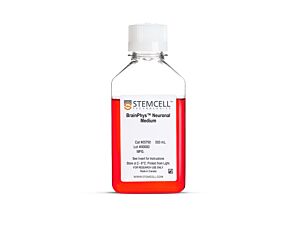
STEMdiff™ Neural Progenitor Medium

Medium for maintenance and expansion of neural progenitor cells derived from human ES and iPS cells

STEMdiff™ Neural Progenitor Medium

Medium for maintenance and expansion of neural progenitor cells derived from human ES and iPS cells

From: 478 USD
Catalog #
(Select a product)
Medium for maintenance and expansion of neural progenitor cells derived from human ES and iPS cells
Add to Wish List

Product Advantages


  • Defined and serum-free

  • Supports expansion of NPCs generated using STEMdiff™ Neural Induction Medium

  • Optimized for efficient expansion of NPCs over multiple passages

  • Preserves NPC multipotency while minimizing spontaneous neuronal differentiation

  • Convenient, user-friendly format and protocol

What's Included

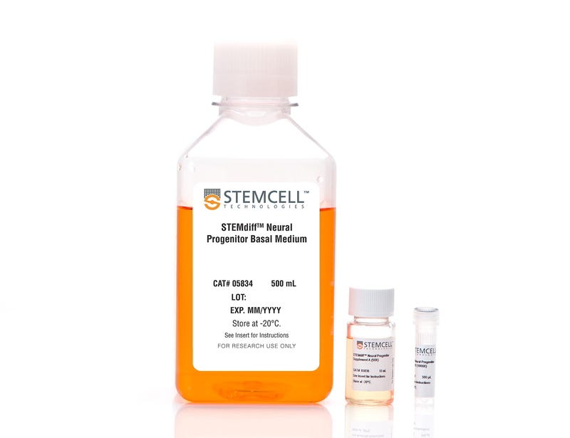
  • STEMdiff™ Neural Progenitor Basal Medium, 500 mL
  • STEMdiff™ Neural Progenitor Supplement A (50X), 10 mL
  • STEMdiff™ Neural Progenitor Supplement B (1000X), 500 µL

Overview

STEMdiff™ Neural Progenitor Medium is a defined and serum-free medium for the expansion of neural progenitor cells (NPCs) derived from human embryonic stem (ES) cells and induced pluripotent stem (iPS) cells using STEMdiff™ Neural Induction Medium (Catalog #05835). NPCs cultured in this medium can be expanded 3-5 fold per passage, and cultured for at least 10 passages, with minimal spontaneous neuronal differentiation.
Subtype
Specialized Media
Cell Type
Neural Cells, PSC-Derived, Neural Stem and Progenitor Cells, Pluripotent Stem Cells
Species
Human
Application
Cell Culture, Expansion
Brand
STEMdiff
Area of Interest
Disease Modeling, Drug Discovery and Toxicity Testing, Neuroscience, Stem Cell Biology
Formulation Category
Serum-Free

Data Figures

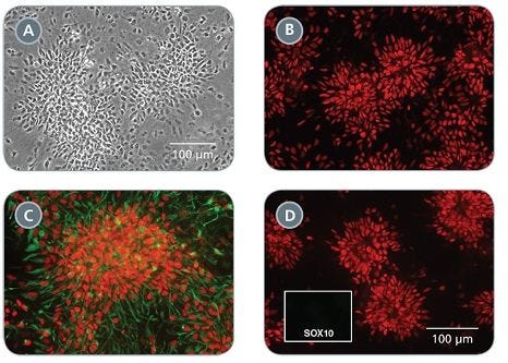
Morphology and Marker Expression of Neural Progenitor Cells Cultured in STEMdiff™ Neural Progenitor Medium

Figure 1. Morphology and Marker Expression of Neural Progenitor Cells Cultured in STEMdiff™ Neural Progenitor Medium

(A) Typical NPC morphology is observed in cultures (shown at day 6 of passage 1). (B-D) NPCs maintained in STEMdiff™ Neural Progenitor Medium express the CNS-type NPC markers PAX6 (B, D, red), SOX1 (C, red) and NESTIN (C, green), but not the neural crest marker SOX10 (D, green, single channel shown in inset). B-D were taken at the same magnification.

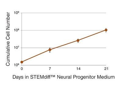
Expansion of Neural Progenitor Cells in STEMdiff™ Neural Progenitor Medium

Figure 2. Expansion of Neural Progenitor Cells in STEMdiff™ Neural Progenitor Medium

NPCs cultured in STEMdiff™ Neural Progenitor Medium can be expanded to generate a large number of cells. Three- to five-fold expansion can be achieved upon each passage. NPCs were derived using STEMdiff™ Neural Induction Medium and passaged once a week on average. n = 6.

Neural Progenitor Cells Cultured in STEMdiff™ Neural Progenitor Medium Show Minimal Spontaneous Neuronal Differentiation

Figure 3. Neural Progenitor Cells Cultured in STEMdiff™ Neural Progenitor Medium Show Minimal Spontaneous Neuronal Differentiation

Passages 1 (A) and 3 (B) of a representative NPC culture maintained in STEMdiff™ Neural Progenitor Medium. Cells were immunolabeled with SOX1 (red) to identify NPCs, and class III β-tubulin (green) to identify neurons. Spontaneous neuronal differentiation is low in NPC cultures maintained in STEMdiff™ Neural Progenitor Medium. A and B were taken at the same magnification.

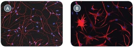
Neural Progenitor Cells Maintained in STEMdiff™ Neural Progenitor Medium can Differentiate into Neurons and Astrocytes

Figure 4. Neural Progenitor Cells Maintained in STEMdiff™ Neural Progenitor Medium can Differentiate into Neurons and Astrocytes

When directed according to published protocols, NPCs can differentiate into neurons (A, class III β-tubulin shown in red) and astrocytes (B, GFAP shown in red). Nuclei are counterstained with DAPI (blue).

Protocols and Documentation

Find supporting information and directions for use in the Product Information Sheet or explore additional protocols below.

Document Type
Product Name
Catalog #
Lot #
Language
Catalog #
05833
Lot #
All
Language
English
Document Type
Technical Manual
Catalog #
05833
Lot #
All
Language
English
Document Type
Safety Data Sheet 1
Catalog #
05833
Lot #
All
Language
English
Document Type
Safety Data Sheet 2
Catalog #
05833
Lot #
All
Language
English
Document Type
Safety Data Sheet 3
Catalog #
05833
Lot #
All
Language
English

Applications

This product is designed for use in the following research area(s) as part of the highlighted workflow stage(s). Explore these workflows to learn more about the other products we offer to support each research area.

Resources and Publications

Publications (15)

Modelling Lyssavirus Infections in Human Stem Cell-Derived Neural Cultures. V. Sundaramoorthy et al. Viruses 2020 mar

Abstract

Rabies is a zoonotic neurological infection caused by lyssavirus that continues to result in devastating loss of human life. Many aspects of rabies pathogenesis in human neurons are not well understood. Lack of appropriate ex-vivo models for studying rabies infection in human neurons has contributed to this knowledge gap. In this study, we utilize advances in stem cell technology to characterize rabies infection in human stem cell-derived neurons. We show key cellular features of rabies infection in our human neural cultures, including upregulation of inflammatory chemokines, lack of neuronal apoptosis, and axonal transmission of viruses in neuronal networks. In addition, we highlight specific differences in cellular pathogenesis between laboratory-adapted and field strain lyssavirus. This study therefore defines the first stem cell-derived ex-vivo model system to study rabies pathogenesis in human neurons. This new model system demonstrates the potential for enabling an increased understanding of molecular mechanisms in human rabies, which could lead to improved control methods.
A Novel Toolkit for Characterizing the Mechanical and Electrical Properties of Engineered Neural Tissues. M. Robinson et al. Biosensors 2019 apr

Abstract

We have designed and validated a set of robust and non-toxic protocols for directly evaluating the properties of engineered neural tissue. These protocols characterize the mechanical properties of engineered neural tissues and measure their electrophysical activity. The protocols obtain elastic moduli of very soft fibrin hydrogel scaffolds and voltage readings from motor neuron cultures. Neurons require soft substrates to differentiate and mature, however measuring the elastic moduli of soft substrates remains difficult to accurately measure using standard protocols such as atomic force microscopy or shear rheology. Here we validate a direct method for acquiring elastic modulus of fibrin using a modified Hertz model for thin films. In this method, spherical indenters are positioned on top of the fibrin samples, generating an indentation depth that is then correlated with elastic modulus. Neurons function by transmitting electrical signals to one another and being able to assess the development of electrical signaling serves is an important verification step when engineering neural tissues. We then validated a protocol wherein the electrical activity of motor neural cultures is measured directly by a voltage sensitive dye and a microplate reader without causing damage to the cells. These protocols provide a non-destructive method for characterizing the mechanical and electrical properties of living spinal cord tissues using novel biosensing methods.
Linc-GALMD1 Regulates Viral Gene Expression in the Chicken. Y. He et al. Frontiers in genetics 2019

Abstract

A rapidly increasing number of reports on dysregulated long intergenic non-coding RNA (lincRNA) expression across numerous types of cancers indicates that aberrant lincRNA expression may be a major contributor to tumorigenesis. Marek's disease (MD) is a T cell lymphoma of chickens induced by Marek's disease virus (MDV). Although we have investigated the roles of lincRNAs in bursa tissue of MDV-infected chickens in previous studies, the molecular mechanisms of lincRNA functions in T cells remain poorly understood. In the present study, Linc-GALMD1 was identified from CD4+ T cells and MSB1 cells, and its expression was significantly downregulated in MD-resistant line of birds in response to MDV challenge. Furthermore, loss-of-function experiments indicated that linc-GALMD1 significantly affected the expression of 290 genes in trans. Through integrated analysis of differentially expressed genes (DEGs) induced by MDV and linc-GALMD1, we found that IGLL1 gene expression levels had a positive correlation with the degree of MD infection and could potentially serve as an indicator for clinical diagnosis of MD. Moreover, an interaction between MDV and linc-GALMD1 was also observed. Accordingly, chicken embryonic fibroblast cells were inoculated with MDV with and without the linc-GALMD1 knockdown, and the data showed that linc-GALMD1 could repress MDV gene expression during the course of MDV infection. These findings uncovered a role of linc-GALMD1 as a viral gene regulator and suggested a function of linc-GALMD1 contributing to tumor suppression by coordinating expression of MDV genes and tumor-related genes and regulating immune responses to MDV infection.