ReLeSR™

cGMP, enzyme-free human pluripotent stem cell selection and passaging reagent

ReLeSR™

cGMP, enzyme-free human pluripotent stem cell selection and passaging reagent
From: 66 USD
Catalog #
(Select a product)
cGMP, enzyme-free human pluripotent stem cell selection and passaging reagent
Add to Wish List

Product Advantages


  • Simplify cell passaging with a straightforward protocol

  • Eliminate the need for manual removal (selection) of differentiated cells

  • Generate cell aggregates without manual scraping

  • Passage cells in flasks and large culture vessels

  • Obtain quality cutures with this gentle, chemically defined, enzyme-free, GMP solution

  • Achieve high expansion of human ES/iPS cells after passaging

Overview

Efficiently dissociate and passage human embryonic stem (ES) or induced pluripotent stem (iPS) cells as aggregates without manual selection or scraping with ReLeSR™. Passaging human ES/iPS cells with ReLeSR™ easily generates optimally-sized aggregates, while eliminating the hassle and variability associated with manual manipulation. By eliminating the need for manual scraping, this enzyme-free reagent enables the use of culture flasks and other closed vessels, facilitating cell culture scale-up and automation. ReLeSR™ is manufactured following relevant cGMPs under a certified quality management system to ensure the highest quality and consistency for reproducible results.

To request a Letter of Authorization (LOA) for ReLeSR’s Drug Master File, click here.
Subtype
Non-Enzymatic
Cell Type
Pluripotent Stem Cells
Species
Human
Application
Cell Culture
Brand
ReLeSR, TeSR
Area of Interest
Stem Cell Biology

Data Figures

Passaging Protocol Comparison

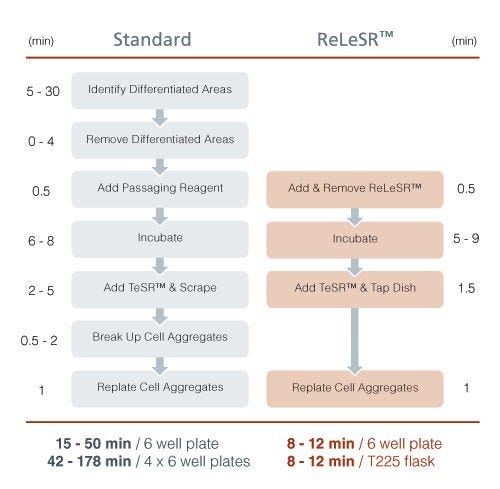
Figure 1. Passaging Protocol Comparison

ReLeSR™ passaging protocol eliminates difficult and time-consuming steps, thereby enabling easy culture scale-up.
Surface area of 4 x 6 well plates (230 cm²) is comparable to that of a T225 flask (225 cm²).
TeSR™ = TeSR™ family media (mTeSR™1, TeSR™2, or TeSR™-E8™).

Selectively Detach Undifferentiated Cells

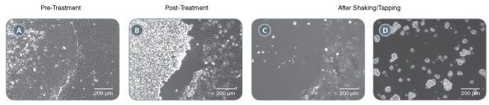
Figure 2. Selectively Detach Undifferentiated Cells

ReLeSR™ selectively detaches undifferentiated cells from pluripotent stem cell cultures without manual selection. Optimally-sized aggregates are generated following shaking/tapping of the cultureware.
(A) An hPSC culture ready for passaging. Note the presence of differentiated cells at the edge of the undifferentiated hPSC colony. (B) Following incubation with ReLeSR™, the undifferentiated hPSC colony starts to lift off of the cultureware. The differentiated cells remain attached to the cultureware. (C) Following shaking/tapping of the cultureware, the undifferentiated cells completely lift off of the cultureware. (D) The undifferentiated hPSC colony is broken up into optimally-sized aggregates for replating.

Rescue Highly Differentiated Cultures

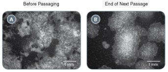
Figure 3. Rescue Highly Differentiated Cultures

Poor quality human pluripotent stem cell cultures containing large proportions of differentiated cells can be rescued by passaging with ReLeSR™. (A) A poor quality hPSC culture containing ~50% undifferentiated cells. (B) Following passaging with ReLeSR™, the differentiated cells have largely been eliminated from the culture, with >90% undifferentiated cells present at the end of the next passage.

Select Putative iPS Cell Clones

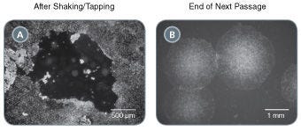
Figure 4. Select Putative iPS Cell Clones

Easily isolate newly generated human iPS cell colonies with ReLeSR™ by selectively detaching undifferentiated cells and leaving non reprogrammed cells behind.
(A) A TeSR™-E7™ reprogramming culture which has been treated with ReLeSR™ to detach the putative iPS cell colony, leaving the non-reprogrammed and differentiated cells behind. (B) Cultures contain a high proportion of undifferentiated cells by the end of the first passage.

Protocols and Documentation

Find supporting information and directions for use in the Product Information Sheet or explore additional protocols below.

Document Type
Product Name
Catalog #
Lot #
Language
Product Name
ReLeSR™
Catalog #
100-0484, 100-0483
Lot #
Lot 1000161526 and higher For 100-0483 | Lot 1000167368 and higher For 100-0484
Language
English
Product Name
ReLeSR™
Catalog #
100-0484, 100-0483
Lot #
Lot 1000161525 and lower For 100-0483 | Lot 1000167367 and lower For 100-0484
Language
English
Document Type
Safety Data Sheet
Product Name
ReLeSR™
Catalog #
100-0484
Lot #
All
Language
English
Document Type
Safety Data Sheet
Product Name
ReLeSR™
Catalog #
100-0483
Lot #
All
Language
English

Applications

This product is designed for use in the following research area(s) as part of the highlighted workflow stage(s). Explore these workflows to learn more about the other products we offer to support each research area.

Resources and Publications

Educational Materials (18)

Publications (194)

Calcium Shock Enables Efficient and Programmable Particle Delivery for Genome Editing Applications N. Vo et al. Advanced Science 2026 Mar

Abstract

Classical intracellular delivery methods such as transfection and transduction are inefficient, particularly with confluent cells and organoids, and lack cell type‐specific programmability. We demonstrate that an innovative methodology called calcium shock (CaSh) dramatically improves particle delivery into single cells, colonies, and organoids, and enables programmable delivery (CaSh‐Pro) into specific cell types within heterocellular populations. Calcium shock works by increasing endocytotic uptake while simultaneously disarming cell‐cell junctions. CaSh‐Pro further incorporates specific molecular targeting agents and amphiphilic peptides for preferential editing of different cell types. Calcium shock improves expression of plasmid, ribonucleoprotein, or adeno‐associated viral vectors with minimal toxicity in intact organoids representing diverse lineages. CaSh and CaSh‐Pro provide simple, versatile protocols for genome editing in complex systems, to enable biological discovery and therapeutic development. Classical transfection and transduction are inefficient, particularly with confluent cells and organoids, and lack cell type‐specific programmability. This study presents calcium shock (CaSh), a method that dramatically improves particle delivery into single cells, colonies, and organoids. CaSh is further utilized to enable programmable delivery (CaSh‐Pro) into specific cell types within heterocellular populations.
Self-Assembly of Human Embryonic-Stem-Cell-Derived Keratinocytes and Fibroblasts into 3D Spheroid Structures for Epidermal Regeneration In Vivo C. Cai et al. Cells 2026 Mar

Abstract

Introduction: Extensive thermal injury remains a formidable clinical challenge, primarily due to the profound deficit of autologous donor skin, which necessitates prolonged hospitalization and escalates healthcare expenditures. While human embryonic stem cells (hESCs) offer a theoretically inexhaustible source for regenerative therapy, optimizing their differentiation and engraftment remains critical for clinical translation. Methods: We used a three-stage protocol to induce the differentiation of hESCs into keratinocytes (KCs). To optimize the delivery of hESC-derived keratinocytes (EKCs), human dermal fibroblasts (HFBs) were utilized to provide essential extracellular matrix (ECM) and microenvironmental support. The two cell types could self-assemble into 3D spheroids. After optimizing the size and cell proportion, these spheroids were subsequently transplanted onto full-thickness dorsal wounds in immunodeficient mice to evaluate their regenerative capacity. Results: hESC-derived keratinocytes exhibited the expression of stage-specific epidermal markers, confirming high differentiation efficiency. In vitro, EKCs demonstrate the capacity to form stratified epidermal structures. By self-assembling into spheres with dermal fibroblasts, the EKCs demonstrated successful engraftment and sustained survival in vivo. The transplantation of these 3D spheroids significantly accelerated wound closure and re-epithelialization compared with controls. Conclusions: This study establishes a robust cell therapy approach characterized by a short preparation cycle with high differentiation efficiency and high transplantation survival rate, offering a novel strategy for the treatment of extensive skin defects.
Modeling neurovascular dysfunction in Alzheimer’s disease using an isogenic brain-chip model A. N. Shen et al. Fluids and Barriers of the CNS 2026 Jan

Abstract

Background: The pathology of Alzheimer’s Disease (AD) is characterized by aggregates of amyloid beta (Aβ) peptides and neurofibrillary tau tangles. Increased blood-brain barrier (BBB) permeability and reduced Aβ clearance, which signal neurovascular dysfunction, have also been proposed as early markers of AD. Despite intense scrutiny, the mechanisms of AD remain elusive and novel treatments that address core symptoms of dementia are limited. New alternative methods (NAMs) aim to develop in-vitro translational models that recapitulate human pathology more accurately than previous models and could contribute to the development of new therapies. Methods: Here, we developed a NAM model of the cortical neurovascular unit (NVU) using brain cells derived from human induced pluripotent stem cells (hiPSCs) from a patient with AD and a healthy individual. Differentiated neurons, astrocytes, pericytes, microglia, and brain-like microvascular endothelial cells were cultured in a microphysiological system to create a brain-chip model to evaluate NVU-related endpoints. Results: Compared to control, AD brain-chips had reduced claudin-5 and ZO-1 expression and increased paracellular permeability. AD brain-chips also had decreased activity of the efflux transporter P-glycoprotein (P-gp), but its expression was unchanged. In AD brain-chips, levels of Aβ42, total tau, and p-tau 181 were decreased in protein lysates from the brain channel, while levels of total tau and p-tau 181 were increased in protein lysates from the vascular channel. Finally, AD brain-chips had increased levels of the proinflammatory markers IL-6 and MCP-1 in effluent from both brain and vascular channels. Conclusion: In this brain-chip model, we showed Aβ-independent NVU dysfunction that was related to neuroinflammation and vascular tau accumulation. This study demonstrates the utility of the brain-chip model to evaluate changes in NVU functions induced by AD-like pathology and highlights donor-specific responses associated with the use of hiPSC-derived models.