ImmunoCult™ Human CD3/CD28/CD2 T Cell Activator

Human T cell activation and expansion reagent

ImmunoCult™ Human CD3/CD28/CD2 T Cell Activator

Human T cell activation and expansion reagent
From: 209 USD
Catalog #
(Select a product)
Human T cell activation and expansion reagent
Add to Wish List

Product Advantages


  • Robust activation and expansion of human T cells without the use of magnetic beads, feeder cells, or antigen

  • Provides a gentle activation stimulus that maintains high viability of activated and expanded T cells

  • Highly stable, filter-sterilized soluble reagent

Overview

Achieve robust activation and expansion of T cells in the absence of magnetic beads, feeder cells, or antigens.

This product’s gentle activation stimulus ensures a high viability of activated T cells, which can be further expanded in ImmunoCult™-XF T Cell Expansion Medium (Catalog #10981) or other media for culturing human T cells. ImmunoCult™ Human CD3/CD28/CD2 T Cell Activator consists of soluble antibody complexes that bind to and cross-link CD3, CD28, and CD2 cell surface ligands, thereby providing the required primary and co-stimulatory signals for T cell activation.

This product is designed for research applications. If you require reagents suitable for use in cell therapy manufacturing, ImmunoCult™ Human CD3/CD28/CD2 T Cell Activators (Catalog #100-0785) are produced under relevant GMPs.
Contains
• Anti-human CD3 monospecific antibody complex
• Anti-human CD28 monospecific antibody complex
• Anti-human CD2 monospecific antibody complex
Subtype
Supplements
Cell Type
T Cells, T Cells, CD4+, T Cells, CD8+
Species
Human
Application
Activation, Cell Culture, Expansion
Brand
ImmunoCult
Area of Interest
Immunology, Cell Therapy Development

Data Figures

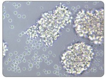
Activated Morphology of Human T Cells Stimulated With ImmunoCult™ Human CD3/CD28/CD2 T Cell Activator

Figure 1. Activated Morphology of Human T Cells Stimulated With ImmunoCult™ Human CD3/CD28/CD2 T Cell Activator

Image of human T cells isolated using the EasySep™ Human T Cell Isolation Kit (Catalog #17951), stimulated with ImmunoCult™ Human CD3/CD28/CD2 T Cell Activator, and cultured in ImmunoCult™-XF T Cell Expansion Medium (Catalog #10981).

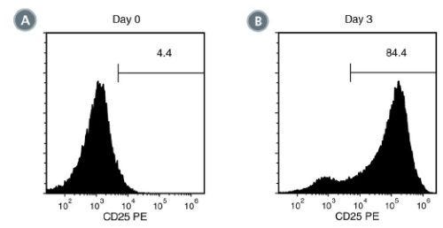
Activation of EasySep™ Isolated Human T Cells Stimulated With ImmunoCult™ Human CD3/CD28/CD2 T Cell Activator

Figure 2. Activation of EasySep™ Isolated Human T Cells Stimulated With ImmunoCult™ Human CD3/CD28/CD2 T Cell Activator

EasySep™-isolated human T cells were stimulated with ImmunoCult™ Human CD3/CD28/CD2 T Cell Activator and cultured in ImmunoCult™-XF T Cell Expansion Medium. Activation of viable CD3+ T cells was assessed by CD25 expression using flow cytometry. On day 0, the frequency of CD25 positive cells was (A) 5.6 ± 2.4% (mean ± SD). Following 3 days of culture, the frequency of CD25 positive cells was (B) 88.8 ± 3.2% (mean ± SD) when stimulated with ImmunoCult™ Human CD3/CD28/CD2 T Cell Activator.

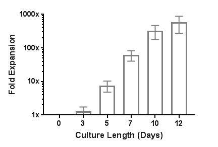
Robust Human T Cell Expansion with ImmunoCult™ Human CD3/CD28/CD2 T Cell Activator

Figure 3. Robust Human T Cell Expansion with ImmunoCult™ Human CD3/CD28/CD2 T Cell Activator

EasySep™-isolated human T cells were expanded over 12 days with ImmunoCult™ Human CD3/CD28/CD2 T Cell Activator in ImmunoCult™-XF T Cell Expansion Medium supplemented with Human Recombinant IL-2. On day 0, 1 x 10^6 EasySep™-isolated human T cells were stimulated with 25 μL of ImmunoCult™ Human CD3/CD28/CD2 T Cell Activator in ImmunoCult™-XF T Cell Expansion Medium supplemented with 10 ng/mL Human Recombinant IL-2. On days 3, 5, 7, and 10, viable cells were counted and fresh medium supplemented with IL-2 was added. No additional ImmunoCult™ Human CD3/CD28/CD2 T Cell Activator was added during the 12-day culture period (mean ± SD in 6 experiments with 3 donors).

Table 1. Comparison of ImmunoCult™ Human T Cell Activation Reagents

Table comparing ImmunoCult™ Human T Cell Activation Reagents

Protocols and Documentation

Find supporting information and directions for use in the Product Information Sheet or explore additional protocols below.

Document Type
Product Name
Catalog #
Lot #
Language
Catalog #
10990, 10970
Lot #
All
Language
English
Document Type
Safety Data Sheet
Catalog #
10990, 10970
Lot #
All
Language
English

Applications

This product is designed for use in the following research area(s) as part of the highlighted workflow stage(s). Explore these workflows to learn more about the other products we offer to support each research area.

Resources and Publications

Educational Materials (21)

Publications (45)

TLR9 Agonists Potentiate Adoptive T-cell Therapy in Cancer through a B-cell-CD2 Costimulatory Axis. A. Ruffin et al. Cancer research 2026 Oct

Abstract

UNLABELLED: Adoptive T-cell transfer (ACT) therapy offers curative potential for some patients with cancer. Toll-like receptor (TLR) agonists improve the efficacy of ACT therapy, and elucidating the underlying mechanism of potency could help determine the best way to maximize the benefits of TLR agonists. In this study, we identified an innate-adaptive circuit in which TLR9-activated B cells augment CD8+ T-cell fitness and antitumor activity through CD2-dependent costimulation. Among multiple TLR agonists tested, class B CpG uniquely programmed murine and human CD8+ T cells for superior effector differentiation, metabolic fitness, and antitumor control. Disruption of CD2 signaling blunted the benefits of TLR9 agonism, including impaired glycolytic capacity and reduced tumor control. Independently, blocking other costimulatory molecules, such as CD86, CD80, CD28, or inducible T-cell costimulator (ICOS), did not impair the antitumor activity of CpG-conditioned T cells. Gain-of-function experiments revealed that CD2 stimulation recapitulated the effect of TLR9 agonism, bolstering the effector function of tumor-infiltrating lymphocytes and chimeric antigen receptor (CAR) T cells. Consistent with these findings, elevated CD2 expression in human tumors correlated with improved overall survival across multiple patient cohorts with cancer, underscoring the clinical importance of this signaling cue. Together, these data uncover a noncanonical B-cell-CD2 costimulation axis through which TLR9 agonists potentiate ACT, revealing a targetable pathway to overcome resistance to cell therapy in solid tumors. SIGNIFICANCE: B cells activated with TLR9 agonist cross-talk with CD8+ T cells via CD2, which enhances their fitness, cytotoxic function, and antitumor activity against solid tumors.
Trophoblast Extracellular Vesicles Rewire Immunity: A Symptom-Relief Avenue for Recessive Dystrophic Epidermolysis Bullosa. N. Hirt et al. Journal of extracellular vesicles 2026 Oct

Abstract

Redirecting immune responses toward tissue repair represents a promising strategy for treating inflammatory and degenerative diseases. Extracellular vesicles (EVs), derived from allogeneic stem cells, have shown potential for modulating proinflammatory immunity. Among these, placental-derived EVs (PEVs) are emerging as candidates for immune-mediated inflammatory disorders due to their role in establishing maternal immune tolerance. However, their applicability beyond pregnancy remains limited by key translational barriers, including the lack of a standardized and scalable source system and need to demonstrate immunomodulatory activity in non-pregnancy allogeneic contexts. In this study, we identify extravillous trophoblasts (EVTs), which play critical role in early placental development, as key source of immunoregulatory PEVs. To overcome donor variability, we employed the HIPEC cell model as established surrogate for primary EVTs and assessed the effects of their derived EVs (EVT-EVs) on circulating monocytes, pivotal mediators of inflammatory responses. High-dimensional molecular profiling revealed that EVT-EVs, similar to EVs derived from first-trimester primary human trophoblast, carry a distinctive anti-inflammatory signature, including a unique miRNA repertoire enriched in immunoregulatory pathways. Integrated proteomic and lipidomic analyses demonstrated that EVT-EVs induce coordinated functional reprograming of monocytes toward an immunomodulatory, inflammation-resolving phenotype. Notably, EVT-EVs also modulated the inflammatory bias of monocytes from patients with recessive dystrophic epidermolysis bullosa (RDEB), a genetic disorder characterized by chronic inflammation, thereby promoting restoration of immune balance. Collectively, these findings identify EVT-EVs as a scalable, off-the-shelf platform with high translational potential for the treatment of chronic inflammatory diseases.
Targeting the ATX-LPA Axis Overcomes TKI Resistance and Immunosuppression in Renal Cell Carcinoma via Dual Inhibition of AKT/mTOR and TBK1/IRF3 Pathways. J. Luo et al. Advanced science (Weinheim, Baden-Wurttemberg, Germany) 2026 Jun

Abstract

BACKGROUND: Therapeutic resistance limits durable survival in advanced/metastatic renal cell carcinoma (RCC) treated with first-line tyrosine kinase inhibitor (TKI) plus immune checkpoint inhibitor (ICI). We sought to define key resistance drivers and actionable targets. METHODS: Integrated RNA sequencing of cabozantinib-resistant RCC cells, lipid metabolomics, and PD-L1 correlation analyses identified ENPP2 as a candidate driver. Its role in TKI resistance and survival signaling was validated by apoptosis, CCK-8, and colony formation assays in vitro and by nude-mouse xenograft models in vivo. ELISA, flow cytometry and tumor cell-T-cell co-culture assays were used to dissect ENPP2-dependent CD8+ T-cell dysfunction. The therapeutic benefit of pharmacologic ATX inhibition combined with standard TKI-ICI regimens was tested in RCC patient-derived xenograft models. RESULTS: The ATX-LPA axis conferred TKI resistance via constitutive AKT/mTOR activation and promoted immune evasion by upregulating PD-L1 through TBK1/IRF3 signaling, thereby impairing intratumoral CD8+ T-cell function. ENPP2 enhanced PD-L1 transcription by facilitating IRF3 nuclear translocation and its direct recruitment to the CD274 promoter. ATX inhibition improved the antitumor efficacy of TKI-ICI therapy in preclinical models. CONCLUSIONS: Targeting the ATX-LPA axis represents a promising strategy to overcome resistance to current TKI-ICI combinations.