Make more informed purchasing decisions with our new product availability and delivery estimate feature, now available on all product pages, in your cart, and during checkout.
Sign In
New to STEMCELL?
Register for an account to quickly and easily purchase products online and for one-click access to all educational content.
Thank you for your interest in this product.
Please provide us with your contact information and your local representative
will contact you with a customized quote. Where appropriate, they can also assist you with a(n):
Estimated delivery time for your area
Product sample or exclusive offer
In-lab demonstration
By submitting this form, you are providing your consent to STEMCELL Technologies Canada Inc. and its subsidiaries and affiliates (“STEMCELL”) to collect and use your information, and send you newsletters and emails in accordance with our privacy policy. Please contact us with any questions that you may have. You can unsubscribe or change your email preferences at any time.
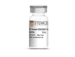
Achieve robust activation and expansion of human T cells for use in clinical applications–without the use of magnetic beads, feeder cells, or antigens.
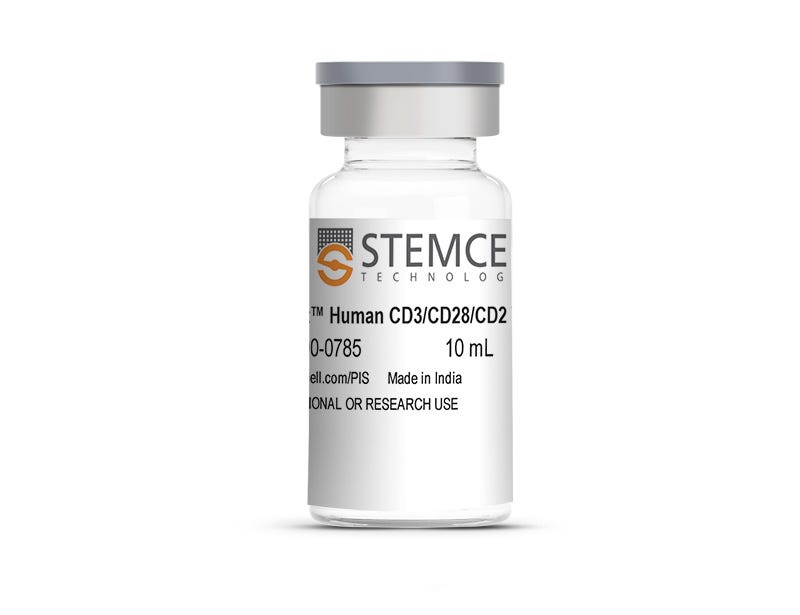
This product’s gentle activation stimulus ensures a high viability of activated T cells, which can be further expanded in ImmunoCult™-XF—a high-performance T cell expansion medium manufactured under relevant cGMP regulations and guidelines. ImmunoCult™ Human CD3/CD28/CD2 T Cell Activator consists of soluble antibody complexes that bind to and cross-link CD3, CD28, and CD2 cell surface ligands, providing the required primary and co-stimulatory signals for T cell culture and activation.
ImmunoCult™ Human CD3/CD28/CD2 T Cell Activator is designed for cell therapy clinical research applications, qualified for use as an ancillary material (AM) following the framework outlined in USP<1043> and/or PH. EUR. 5.2.12. STEMCELL can work with you to qualify this reagent as an AM under an approved Investigational New Drug (IND) application, Biological Licensing Application (BLA), or Clinical Trial Application (CTA). Learn more about how we can support your regulatory needs here.
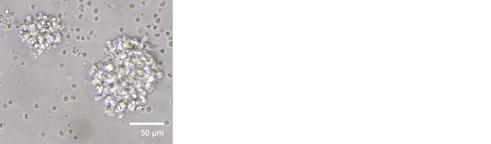
Figure 1. Morphology of Activated Human T Cells Stimulated with ImmunoCult™ Human CD3/CD28/CD2 T Cell Activator
A clustered morphology is seen in activated human T cells. Cells were isolated using EasySep™ Human T Cell Isolation Kit, stimulated with ImmunoCult™ Human CD3/CD28/CD2 T Cell Activator for 3 days in ImmunoCult™-XF supplemented with recombinant human interleukin-2 (rhIL-2).
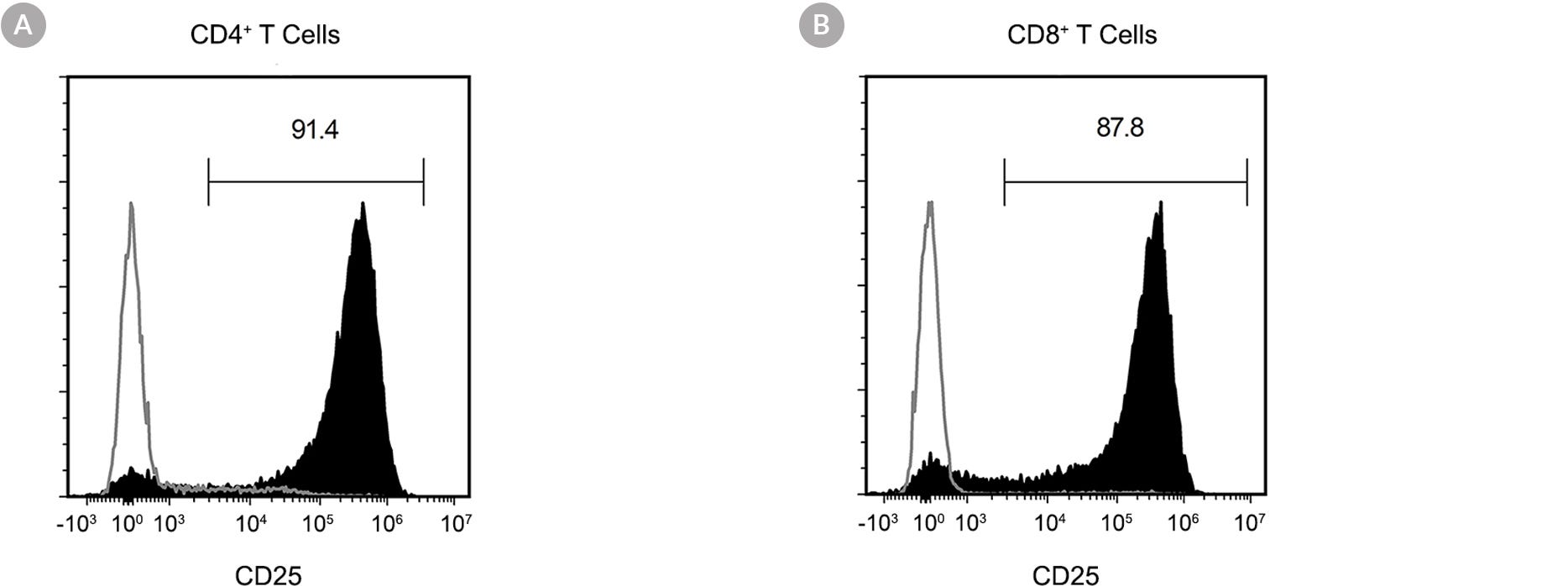
Figure 2. Activation of Human T Cells Stimulated with ImmunoCult™ Human CD3/CD28/CD2 Activator.
Human T cells were stimulated with ImmunoCult™ Human CD3/CD28/CD2 T Cell Activator and cultured in ImmunoCult™-XF. Activation of viable CD4+ and CD8+ T cells were assessed by CD25 expression, using flow cytometry. Following 3 days of culture, the frequency of CD25-positive cells was (A) 91.4% for CD4+ T cells and (B) 87.8% for CD8+ T cells. The gray line depicts day 3 CD4+ and CD8+ T cells cultured without ImmunoCult™ Human CD3/CD28/CD2 T Cell Activator.
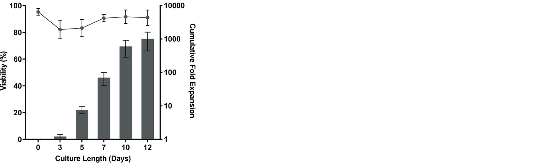
Figure 3. Robust Human T Cell Expansion and High Viability Achieved Using ImmunoCult™ Human CD3/CD28/CD2 T Cell Activator
Human T cells were expanded over 12 days with ImmunoCult™ Human CD3/CD28/CD2 T Cell Activator in ImmunoCult™-XF supplemented with rhIL-2. On day 0, 1 x 10^6 isolated human T cells were stimulated with 25 μL of ImmunoCult™ Human CD3/CD28/CD2 T Cell Activator in ImmunoCult™-XF supplemented with rhIL-2.No additional ImmunoCult™ Human CD3/CD28/CD2 T Cell Activator was added during the 12-day culture period (mean ± SD in 3 experiments with 7 donors).
This product is designed for use in the following research area(s) as part
of the highlighted workflow stage(s). Explore these workflows to learn more about the other products we
offer to support each research area.
Pharmacological Inhibition of MALT1 Ameliorates Autoimmune Pathogenesis and Can Be Uncoupled From Effects on Regulatory T-Cells.
S. Biswas et al.
Frontiers in immunology 2022
Abstract
MALT1 forms part of a central signaling node downstream of immunoreceptor tyrosine-based activation motif (ITAM)-containing receptors, across a broad range of immune cell subsets, and regulates NF-$\kappa$B driven transcriptional responses via dual scaffolding-protease activity. Allosteric inhibition of MALT1 activity has demonstrated benefit in animal models of inflammation. However, development of MALT1 inhibitors to treat autoimmune and inflammatory diseases (A&ID) has been hindered by reports linking MALT1 inhibition and genetic loss-of-function to reductions in regulatory T-cell (Treg) numbers and development of auto-inflammatory syndromes. Using an allosteric MALT1 inhibitor, we investigated the consequence of pharmacological inhibition of MALT1 on proinflammatory cells compared to regulatory T-cells. Consistent with its known role in ITAM-driven responses, MALT1 inhibition suppressed proinflammatory cytokine production from activated human T-cells and monocyte-derived macrophages, and attenuated B-cell proliferation. Oral administration of a MALT1 inhibitor reduced disease severity and synovial cytokine production in a rat collagen-induced arthritis model. Interestingly, reduction in splenic Treg numbers was less pronounced in the context of inflammation compared with na{\{i}}ve animals. Additionally in the context of the disease model we observed an uncoupling of anti-inflammatory effects of MALT1 inhibition from Treg reduction with lower systemic concentrations of inhibitor needed to reduce disease severity compared to that required to reduce Treg numbers. MALT1 inhibition did not affect suppressive function of human Tregs in vitro. These data indicate that anti-inflammatory efficacy can be achieved with MALT1 inhibition without impacting the number or function of Tregs further supporting the potential of MALT1 inhibition in the treatment of autoimmune disease."
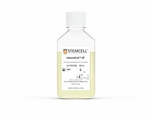
cGMP, serum-free, and xeno-free medium for the expansion of human T cells
Item added to your cart
ImmunoCult™ Human CD3/CD28/CD2 T Cell Activator
Quality Statement:
THIS PRODUCT IS MANUFACTURED AND TESTED FOLLOWING RELEVANT CGMPs UNDER A CERTIFIED QUALITY MANAGEMENT SYSTEM. PRODUCT IS FOR INVESTIGATIONAL OR RESEARCH USE. NOT INTENDED FOR HUMAN OR ANIMAL DIAGNOSTIC OR THERAPEUTIC USES UNLESS OTHERWISE STATED. FOR ADDITIONAL INFORMATION ON QUALITY AT STEMCELL, REFER TO WWW.STEMCELL.COM/COMPLIANCE.